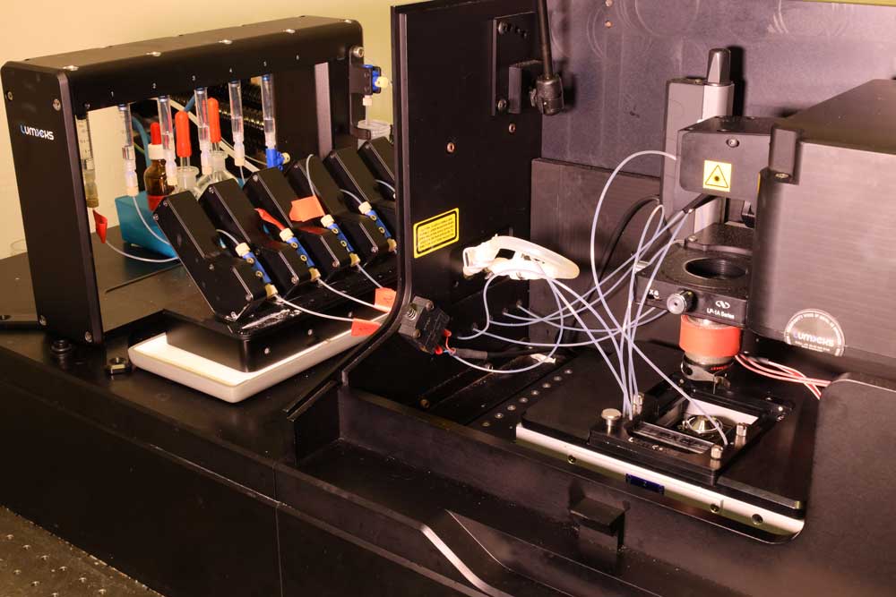
The multicolor confocal fluorescence imaging allows the visualization of biological processes such as protein binding kinetics on DNA. These interactions of fluorescently labeled molecules can be visualized by kymographs, a graph in which the y-axis shows position and the x-axis shows time. The resulting kymograph unveils the number, position, diffusion, and (un-)binding kinetics of the proteins along the DNA. With its fast 1D scanning capabilities, the confocal C-Trap is suitable for constructs such as DNA or other filaments. Super-resolution imaging is possible via 1D STED. The system can be run in a time-tagged mode, enabling fluorescence correlation spectroscopy (FCS) measurements.
Instrument Configuration
- Max. number of traps: 4
- Force resolution (x,y): < 0.1 pN @100 Hz
- Bead displacement resolution using force signal: < 0.3 nm @ 100 Hz
- Bead displacement resolution using live bright-field bead tracking: < 3 nm @ 100 Hz
- Brightfield field of view (x,y): 111 µm x 70 µm (full ROI), pixel size = 115 nm
- Brightfield camera frame rate: ~150 FPS (full ROI, depending on settings)
- Confocal field of view (x,y): 55 µm x 40 µm (full ROI), pixel size = 100 nm (standard, 20 nm for STED)
- Confocal frame rate:
- 2D scan: 0.04 fps (full ROI) to 0.3 fps (smaller, realistic ROI)
- Line scan: 17 fps (full line in x) to 30 fps (smaller, realistic line)
- Available excitation laser lines: 488 nm / 561 nm / 639 nm
- Available depletion laser line: 767 nm
- Available detectors: 3 APDs
- Detection filters: 525/40, 600/50, 680/42
- Microfluidics unit: advanced u-Flux microfluidics with automated valves, 5 channels + 1 outlet, pressure 0.1 - 2.5 bar, minimal reservoir volume 250 ul per channel
- Environmental control: temperature control of condenser and objective >28 °C +- 0.05 °C
- Maximal thickness of flow cell or sample chamber: 1.3 mm
Objective
- CFI Plan Apo VC 60XC WI, NA = 1.2 (water immersion, Nikon)
Condenser