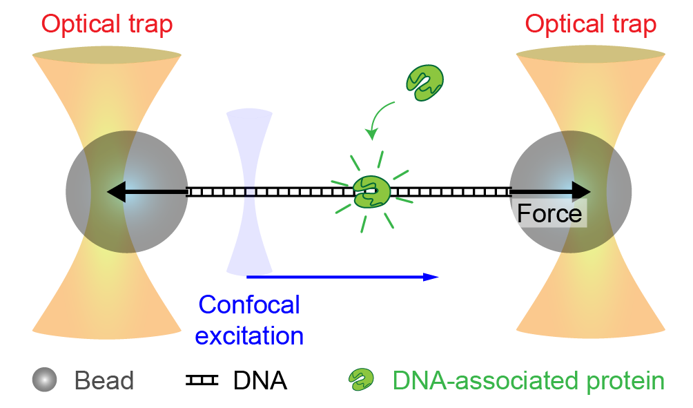
Optical tweezers are advanced scientific instruments that use lasers to trap and manipulate microscopic particles, such as cells or beads. Developed by Arthur Ashkin in 1970, optical tweezers employ the gradient force created by a tightly focused infrared laser to hold particles in three-dimensional space. These particles can be coated to stick to a variety of biomolecules, such as proteins, cytoskeleton filaments, DNA, or RNA. As a result, it is possible to measure and apply forces in the pN range to these molecules during e.g. protein-protein/-DNA interactions, protein folding, phase separation or cytoskeletal transport. Displacements by e.g. conformational changes can be measured in the nm range.
Available systems:
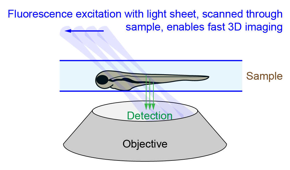
Swept confocally-aligned planar excitation (SCAPE) systems are light sheet fluorescence microscopes that use a thin sheet of light to illuminate a specific plane of a specimen to reduce phototoxicity and photobleaching while enabling fast volumetric imaging with excellent optical sectioning. Unlike traditional light sheet microscopes, SCAPE employs only a single objective lens for both illumination and detection so large samples such as multi-well slides / plates can easily mounted. For 3D volumetric imaging the illumination light sheet is scanned through the sample using a galvanometric mirror to achieve acquisition rates of >10 volumes per second. Alternatively, larger samples scan be scanned by moving the sample through the light sheet using a motorized stage. The acquired data can be deskewed using a GPU-accelerated algorithm. The microscope is best suited for small-to-medium-sized structures such as cell cultures, organoids, or small embryos at high speed for fast temporal resolution or high throughput.
Available systems:
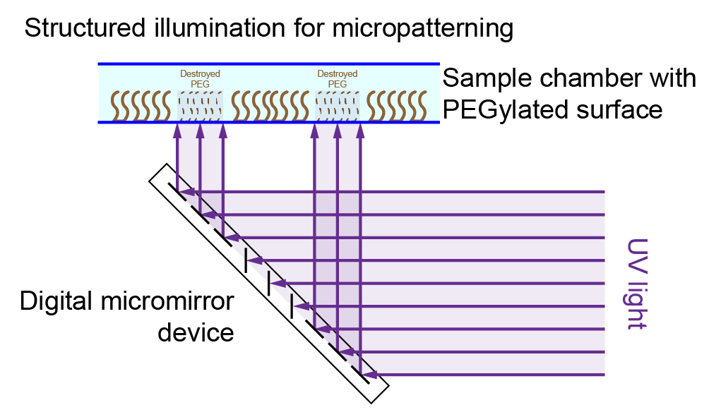
The Primo micropatterning system is a widefield microscope equipped with an Alveolé Primo-1 digital micromirror device (DMD) for illuminating the field of view with a pattern. By illuminating the sample with UV light, the system allows for creating patterns of surface coating on glass substrates and TEM grids for electron microscopy (e.g. local PEG passivation). In addition, the system is capable to generate 3D hydrogel structures on surfaces.
Available systems:
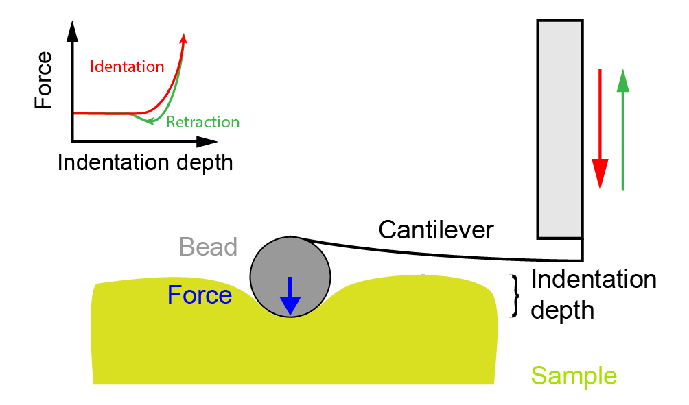
In nanoindentation measurements, cantilevers with micrometer-sized spheres are pushed into a soft sample like a gel, a cell, organoids, or tissues. The probe’s indentation into the sample as a function of corresponding force are measured. Quasi-static indentation and relaxation measurements give insight into material properties like Young’s modulus. Dynamic indentations with sinusoidal modulation enable to measure frequency-dependent material properties like storage and loss moduli.
Available systems: