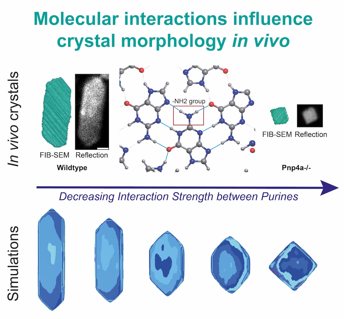
Purine Molecular Interactions Determine Anisotropic Shape of Zebrafish Biogenic Crystals
Across phyla, many organisms self-organize crystals, for functions like vision, pigmentation, and metabolite storage. In zebrafish, a vertebrate known for its crystal-based color patterns, iridophores concentrate purines in membrane-bound organelles, the iridosomes. Inside these vesicles, crystals assemble into large, flat, and thin hexagons following unknown mechanisms that defy typical thermodynamic interactions. Here, we investigate the development of zebrafish iridosomal crystals by using live imaging, cryoFIB-SEM, and novel morphometric analysis pipelines. In doing so, we find that crystal growth predominantly occurs along the b-crystallographic axis, producing their characteristic anisotropic shape. By performing comparative genetic analyses in vivo and reproducing such conditions in silico, we uncover that the zebrafish crystals' in-plane hydrogen bond molecular structure is the main determinant for the observed crystal anisotropy. Macroscopically, the b-axis anisotropy is controlled by the ratio of guanine-to-hypoxanthine in the iridosome, without affecting the other axes. At the atomic level, the extent of the (100) facet anisotropy depends entirely on the type, number, and strength of molecular H-bonds within the crystal lattice. Mechanistically, our work shows that purine diversity and availability inside the zebrafish iridosome is key to form an anisotropic crystal lattice, leading to the observed functional crystal shapes.
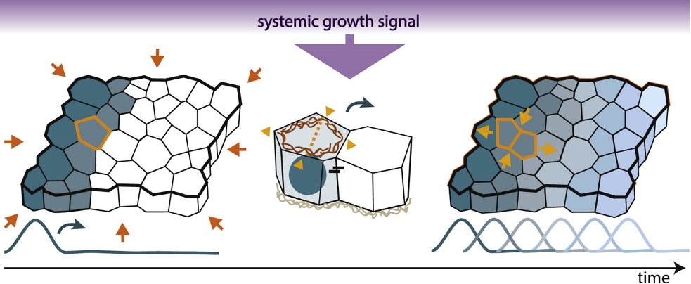
Growth across scales: Dynamic signaling impacts tissue size and shape
Organ and tissue growth result from an integration of biophysical communication across biological scales, both in time and space. In this review, we highlight new insight into the dynamic connections between control mechanisms operating at different length scales. First, we consider how the dynamics of chemical and electrical signaling in the shape of gradients or waves affect spatiotemporal signal interpretation. Then, we discuss the mechanics underlying dynamic cell behavior during oriented tissue growth, followed by the connections between signaling at the tissue and organismal levels.
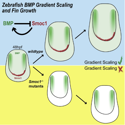
BMP Signaling Gradient Scaling in the Zebrafish Pectoral Fin
Secreted growth factors can act as morphogens that form spatial concentration gradients in developing organs, thereby controlling growth and patterning. For some morphogens, adaptation of the gradients to tissue size allows morphological patterns to remain proportioned as the organs grow. In the zebrafish pectoral fin, we found that BMP signaling forms a two-dimensional gradient. The length of the gradient scales with tissue length and its amplitude increases with fin size according to a power-law. Gradient scaling and amplitude power-laws are signatures of growth control by time derivatives of morphogenetic signaling: cell division correlates with the fold change over time of the cellular signaling levels. We show that Smoc1 regulates BMP gradient scaling and growth in the fin. Smoc1 scales the gradient by means of a feedback loop: Smoc1 is a BMP agonist and BMP signaling represses Smoc1 expression. Our work uncovers a layer of morphogen regulation during vertebrate appendage development.
Jinghui Liu*, Elisa Nerli*, Charlie Duclut, Amit S Vishen, Naomi Berbee, Sylvia Kaufmann, Cesar Ponce, Aristides B. Arrenberg, Frank Jülicher#, Rita Mateus# Injury-induced electrochemical coupling triggers organ growth. Sci Adv, 12(6) Art. No. 687 (2026)
Open Access DOI
Organ injury triggers nonneuronal electric currents essential for regeneration. However, the mechanisms by which electrical signals are generated, sensed, and transmitted upon damage to promote organ growth remain unclear. Here, we uncover that organ repair relies on dynamic electrochemical coupling between membrane potential depolarization and intracellular signaling, essential to activate cell proliferation. By subsecond live imaging of locally injured zebrafish larval fins, we identify events across time and space: a millisecond, long-range, membrane depolarization gradient, followed by second-persistent intracellular calcium responses. In the subsequent hour, voltage sensing phosphatase senses the injury-driven membrane potential change and autonomously translates the electric signal intracellularly, promoting tissue-wide cell proliferation. Connecting these dynamics with an electrodiffusive model showed that ionic fluxes and electric potential become coupled in the fin's interstitial space, enabling organ-wide signal spreading. Our work reveals the coupling between fast electrical signals and slower intracellular signaling, ensuring complete organ recovery.
2025
Jannik Rothkegel, Sylvia Kaufmann, Michaela Wilsch-Bräuninger, Catarina Lopes, Rita Mateus Purine Molecular Interactions Determine Anisotropic Shape of Zebrafish Biogenic Crystals. Small Methods, 9(9) Art. No. e01956 (2025)
Open Access DOI
Across phyla, many organisms self-organize crystals, for functions like vision, pigmentation, and metabolite storage. In zebrafish, a vertebrate known for its crystal-based color patterns, iridophores concentrate purines in membrane-bound organelles, the iridosomes. Inside these vesicles, crystals assemble into large, flat, and thin hexagons following unknown mechanisms that defy typical thermodynamic interactions. Here, we investigate the development of zebrafish iridosomal crystals by using live imaging, cryoFIB-SEM, and novel morphometric analysis pipelines. In doing so, we find that crystal growth predominantly occurs along the b-crystallographic axis, producing their characteristic anisotropic shape. By performing comparative genetic analyses in vivo and reproducing such conditions in silico, we uncover that the zebrafish crystals' in-plane hydrogen bond molecular structure is the main determinant for the observed crystal anisotropy. Macroscopically, the b-axis anisotropy is controlled by the ratio of guanine-to-hypoxanthine in the iridosome, without affecting the other axes. At the atomic level, the extent of the (100) facet anisotropy depends entirely on the type, number, and strength of molecular H-bonds within the crystal lattice. Mechanistically, our work shows that purine diversity and availability inside the zebrafish iridosome is key to form an anisotropic crystal lattice, leading to the observed functional crystal shapes.
Lucas Ribas, Rita Mateus Start-Shape-Stop: Cell communication mechanisms controlling organ size. Semin Cell Dev Biol, 174 Art. No. 103641 (2025)
Open Access DOI
Accurate growth control is critical for the achievement of proportional organs during animal development and repair processes. Either extra or deficient growth rates lead to organ functional impairment. The understanding of how organs acquire, recover, and fine-tune their final size has been a long-lasting biological problem. How do organs measure their current size? This review is centered on this question through the lens of the physical properties governing cell communication mechanisms. In particular, we highlight and discuss new insight into the dynamic connections between several cellular control mechanisms that operate at different timescales to regulate organ growth and morphogenesis.
Rachael Deis, Tali Lerer-Goldshtein, Olha Baiko, Zohar Eyal, Dolev Brenman-Begin, Moshe Goldsmith, Sylvia Kaufmann, Uwe Heinig, Yonghui Dong, Sofya Lushchekina, Neta Varsano, Tsviya Olender, Meital Kupervaser, Ziv Porat, Smadar Levin-Zaidman, Iddo Pinkas, Rita Mateus, Dvir Gur Genetic control over biogenic crystal morphogenesis in zebrafish. Nat Chem Biol, 21(3) 383-392 (2025)
Open Access DOI
Organisms evolve mechanisms that regulate the properties of biogenic crystals to support a wide range of functions, from vision and camouflage to communication and thermal regulation. Yet, the mechanism underlying the formation of diverse intracellular crystals remains enigmatic. Here we unravel the biochemical control over crystal morphogenesis in zebrafish iridophores. We show that the chemical composition of the crystals determines their shape, particularly through the ratio between the nucleobases guanine and hypoxanthine. We reveal that these variations in composition are genetically controlled through tissue-specific expression of specialized paralogs, which exhibit remarkable substrate selectivity. This orchestrated combination grants the organism with the capacity to generate a broad spectrum of crystal morphologies. Overall, our findings suggest a mechanism for the morphological and functional diversity of biogenic crystals and may, thus, inspire the development of genetically designed biomaterials and medical therapeutics.
2024
Bethan Clark, Aaron Hickey, Aleksandra Marconi, Bettina Fischer, Joel Elkin, Rita Mateus, M Emília Santos Developmental plasticity and variability in the formation of egg-spots, a pigmentation ornament in the cichlid Astatotilapia calliptera. Evol Dev, 26(3) Art. No. e12475 (2024)
Open Access DOI
Vertebrate pigmentation patterns are highly diverse, yet we have a limited understanding of how evolutionary changes to genetic, cellular, and developmental mechanisms generate variation. To address this, we examine the formation of a sexually-selected male ornament exhibiting inter- and intraspecific variation, the egg-spot pattern, consisting of circular yellow-orange markings on the male anal fins of haplochromine cichlid fishes. We focus on Astatotilapia calliptera, the ancestor-type species of the Malawi cichlid adaptive radiation of over 850 species. We identify a key role for iridophores in initializing egg-spot aggregations composed of iridophore-xanthophore associations. Despite adult sexual dimorphism, aggregations initially form in both males and females, with development only diverging between the sexes at later stages. Unexpectedly, we found that the timing of egg-spot initialization is plastic. The earlier individuals are socially isolated, the earlier the aggregations form, with iridophores being the cell type that responds to changes to the social environment. Furthermore, we observe apparent competitive interactions between adjacent egg-spot aggregations, which strongly suggests that egg-spot patterning results mostly from cell-autonomous cellular interactions. Together, these results demonstrate that A. calliptera egg-spot development is an exciting model for investigating pigment pattern formation at the cellular level in a system with developmental plasticity, sexual dimorphism, and intraspecific variation. As A. calliptera represents the ancestral bauplan for egg-spots, these findings provide a baseline for informed comparisons across the incredibly diverse Malawi cichlid radiation.
2021
Rita Mateus#, Jana Fuhrmann, Natalie Dye# Growth across scales: Dynamic signaling impacts tissue size and shape. Curr Opin Cell Biol, 73 50-57 (2021)
Open Access DOI
Organ and tissue growth result from an integration of biophysical communication across biological scales, both in time and space. In this review, we highlight new insight into the dynamic connections between control mechanisms operating at different length scales. First, we consider how the dynamics of chemical and electrical signaling in the shape of gradients or waves affect spatiotemporal signal interpretation. Then, we discuss the mechanics underlying dynamic cell behavior during oriented tissue growth, followed by the connections between signaling at the tissue and organismal levels.
2020
Rita Mateus*, Laurent Holtzer*, Carole Seum, Zena Hadjivasiliou, Marine Dubois, Frank Jülicher, Marcos Gonzalez-Gaitan BMP Signaling Gradient Scaling in the Zebrafish Pectoral Fin. Cell Rep, 30(12) 4292-4302 (2020)
Open Access DOI
Secreted growth factors can act as morphogens that form spatial concentration gradients in developing organs, thereby controlling growth and patterning. For some morphogens, adaptation of the gradients to tissue size allows morphological patterns to remain proportioned as the organs grow. In the zebrafish pectoral fin, we found that BMP signaling forms a two-dimensional gradient. The length of the gradient scales with tissue length and its amplitude increases with fin size according to a power-law. Gradient scaling and amplitude power-laws are signatures of growth control by time derivatives of morphogenetic signaling: cell division correlates with the fold change over time of the cellular signaling levels. We show that Smoc1 regulates BMP gradient scaling and growth in the fin. Smoc1 scales the gradient by means of a feedback loop: Smoc1 is a BMP agonist and BMP signaling represses Smoc1 expression. Our work uncovers a layer of morphogen regulation during vertebrate appendage development.
2019
Ana S Brandão, Anabela Bensimon-Brito#, Raquel Lourenço, Jorge Borbinha, Ana Rosa Soares, Rita Mateus, António Jacinto# Yap induces osteoblast differentiation by modulating Bmp signalling during zebrafish caudal fin regeneration. J Cell Sci, 132(22) Art. No. jcs231993 (2019)
DOI
Osteoblast differentiation is a key process for bone homeostasis and repair. Multiple signalling pathways have been associated with osteoblast differentiation, yet much remains unknown on how this process is regulated in vivo Previous studies have proposed that the Hippo pathway transcriptional co-activators YAP and TAZ (also known as YAP1 and WWTR1, respectively) maintain progenitor stemness and inhibit terminal differentiation of osteoblasts, whereas others suggest they potentiate osteoblast differentiation and bone formation. Here, we use zebrafish caudal fin regeneration as a model to clarify how the Hippo pathway regulates de novo bone formation and osteoblast differentiation. We demonstrate that Yap inhibition leads to accumulation of osteoprogenitors and prevents osteoblast differentiation in a cell non-autonomous manner. This effect correlates with a severe impairment of Bmp signalling in osteoblasts, likely by suppressing the expression of the ligand bmp2a in the surrounding mesenchymal cells. Overall, our findings provide a new mechanism of bone formation through the Hippo-Yap pathway, integrating Yap in the signalling cascade that governs osteoprogenitor maintenance and subsequent differentiation during zebrafish caudal fin regeneration.
2015
Rita Mateus, Raquel Lourenço, Yi Fang, Goncalo Brito, Ana Farinho, Fábio Valério, Antonio Jacinto Control of tissue growth by Yap relies on cell density and F-actin in zebrafish fin regeneration. Development, 142(16) 2752-2763 (2015)
DOI
Caudal fin regeneration is characterized by a proliferation boost in the mesenchymal blastema that is controlled precisely in time and space. This allows a gradual and robust restoration of original fin size. However, how this is established and regulated is not well understood. Here, we report that Yap, the Hippo pathway effector, is a chief player in this process: functionally manipulating Yap during regeneration dramatically affects cell proliferation and expression of key signaling pathways, impacting regenerative growth. The intracellular location of Yap is tightly associated with different cell densities along the blastema proximal-distal axis, which correlate with alterations in cell morphology, cytoskeleton and cell-cell contacts in a gradient-like manner. Importantly, Yap inactivation occurs in high cell density areas, conditional to F-actin distribution and polymerization. We propose that Yap is essential for fin regeneration and that its function is dependent on mechanical tension, conferred by a balancing act of cell density and cytoskeleton activity.
2012
Rita Mateus, Telmo Pereira, Sara Sousa, Joana Esteves de Lima, Susana Pascoal, Leonor Saúde, Antonio Jacinto In vivo cell and tissue dynamics underlying zebrafish fin fold regeneration. PLoS ONE, 7(12) Art. No. e51766 (2012)
Open Access DOI
Zebrafish (Danio rerio) has a remarkable capacity to regenerate many organs and tissues. During larval stages the fin fold allows the possibility of performing long time-lapse imaging making this system very appealing to study the relationships between tissue movements, cell migration and proliferation necessary for the regeneration process.