“We aim to reveal physical principles and develop physical concepts that underly the dynamic self-organization of living matter.”
In the Grill Lab at the MPI-CBG in Dresden, we investigate the fundamental physical principles that drive the dynamic self-organization of living systems. Our research seeks to understand how mechanical forces, active processes, and emergent physical behaviors shape the architecture of cells, tissues, and embryos. By combining quantitative experiments with theoretical physics, we reveal how structure and function emerge at the mesoscale, from molecular assemblies to multicellular organization.
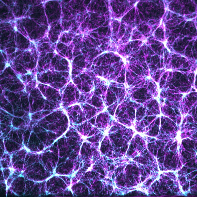
We study how physical principles shape life. We pursue an active matter physics approach to investigate how the actomyosin cytoskeleton inside the nematode Caenorhabditis elegans generates the forces that polarize the zygote, and the torques that drive left-right symmetry breaking. We use optical tweezers to investigate collective protein-DNA interactions and their dependence on DNA sequence. Through this, we work to advance our understanding of the physical concepts and principles that govern the dynamic self-organization of living matter, to shed light on how living systems build and structure themselves over time.
Projects
What mechanisms drive morphogenesis?
We explore how asymmetries arise during development through the mechanical activity of cytoskeletal components. We focus on the actomyosin cortical layer, and combine active matter theory with experiments to uncover the physical mechanisms by which generation of active tension and chiral active torque guide symmetry breaking and body axis establishment in the early C. elegans embryo. In the past we have investigated mechanisms of guided mechano-chemical self-organization in the context of cell polarity establishment in the C. elegans zygote. A current focus is the physical basis of chiral morphogenesis and the establishment of the left-right body axis via active torque generation in the actomyosin cortical layer. For this, we are also investigating tissue-scale torque generation in developing quail embryos, to shed light on the process of left-right body axis formation in birds.
Other areas of focus include C. elegans eggshell formation and hydraulic decision making processes in the syncytial C. elegans gonad.
How is the nucleus organized?
Our lab explores the physical principles that govern the sequence-dependent organization of protein-DNA assemblies. By integrating theory with in vitro assays using optical tweezers, we uncovered two distinct mechanisms of collective protein-DNA interactions: transcription factor prewetting via a DNA-mediated collective surface phase transitions, and protein-DNA co-condensation via a globular collapse of DNA. These physical mechanisms participate in mesoscale structural organization of the nucleus, and offer novel perspectives for the regulation of gene expression.
In collaboration with the von Appen lab at MPI-CBG we are now extending these studies to the nuclear periphery. Here, we identified an unconventional DNA stiffening effect based on BAF and LEM2, together forming a load-bearing, mesoscale surface hydrogel that reinforces the nuclear periphery to provide genome integrity.
What are the physical mechanisms of life?
Theory, in particular active matter physics theory, is integral to our approach, providing a quantitative framework to uncover the physical mechanisms driving self-organization in living systems. We develop continuum and coarse-grained descriptions of active living matter, to describe dynamics, force generation, and symmetry breaking across scales.
Teije C Middelkoop#, Jonas Neipel, Caitlin E Cornell, Ronald Naumann, Lokesh G Pimpale, Frank Jülicher#, Stephan W. Grill# A cytokinetic ring-driven cell rotation achieves Hertwig's rule in early development. Proc Natl Acad Sci U.S.A., 121(25) Art. No. e2318838121 (2024)
Open Access DOI
Hertwig's rule states that cells divide along their longest axis, usually driven by forces acting on the mitotic spindle. Here, we show that in contrast to this rule, microtubule-based pulling forces in early Caenorhabditis elegans embryos align the spindle with the short axis of the cell. We combine theory with experiments to reveal that in order to correct this misalignment, inward forces generated by the constricting cytokinetic ring rotate the entire cell until the spindle is aligned with the cell's long axis. Experiments with slightly compressed mouse zygotes indicate that this cytokinetic ring-driven mechanism of ensuring Hertwig's rule is general for cells capable of rotating inside a confining shell, a scenario that applies to early cell divisions of many systems.
Victoria T Yan*, Arjun Narayanan*, Tina Wiegand, Frank Jülicher#, Stephan W. Grill# A condensate dynamic instability orchestrates actomyosin cortex activation. Nature, 609(7927) 597-604 (2022)
Open Access DOI
A key event at the onset of development is the activation of a contractile actomyosin cortex during the oocyte-to-embryo transition1-3. Here we report on the discovery that, in Caenorhabditis elegans oocytes, actomyosin cortex activation is supported by the emergence of thousands of short-lived protein condensates rich in F-actin, N-WASP and the ARP2/3 complex4-8 that form an active micro-emulsion. A phase portrait analysis of the dynamics of individual cortical condensates reveals that condensates initially grow and then transition to disassembly before dissolving completely. We find that, in contrast to condensate growth through diffusion9, the growth dynamics of cortical condensates are chemically driven. Notably, the associated chemical reactions obey mass action kinetics that govern both composition and size. We suggest that the resultant condensate dynamic instability10 suppresses coarsening of the active micro-emulsion11, ensures reaction kinetics that are independent of condensate size and prevents runaway F-actin nucleation during the formation of the first cortical actin meshwork.
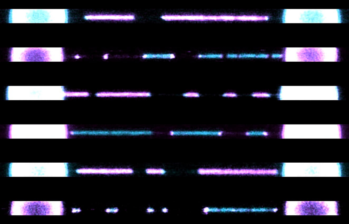
Jose A. Morin*, Sina Wittmann*, Sandeep Choubey*, Adam Klosin, Stefan Golfier, Anthony A. Hyman#, Frank Jülicher#, Stephan W. Grill# Sequence-dependent surface condensation of a pioneer transcription factor on DNA. Nat Phys, 18(3) 271-276 (2022)
Open Access DOI
Biomolecular condensates are dense assemblies of proteins that form distinct biochemical compartments without being surrounded by a membrane. Some, such as P granules and stress granules, behave as droplets and contain many millions of molecules. Others, such as transcriptional condensates that form on the surface of DNA, are small and contain thousands of molecules. The physics behind the formation of small condensates on DNA surfaces is still under discussion. Here we investigate the nature of transcription factor condensates using the pioneer transcription factor Kruppel-like factor 4 (Klf4). We show that Klf4 can phase separate on its own at high concentrations, but at low concentrations, Klf4 only forms condensates on DNA. Using optical tweezers, we demonstrate that these Klf4 condensates form on DNA as a type of surface condensation. This surface condensation involves a switch-like transition from a thin adsorbed layer to a thick condensed layer, which shows hallmarks of a prewetting transition. The localization of condensates on DNA correlates with sequence, suggesting that the condensate formation of Klf4 on DNA is a sequence-dependent form of surface condensation. Prewetting together with sequence specificity can explain the size and position control of surface condensates. We speculate that a prewetting transition of pioneer transcription factors on DNA underlies the formation and positioning of transcriptional condensates and provides robustness to transcriptional regulation.
A DNA-binding protein condenses on DNA via a switch-like transition. Surface condensation occurs at preferential DNA locations suggesting collective sequence readout and enabling sequence-specificity robustness with respect to protein concentration.
Nicolas T Chartier*, Arghyadip Mukherjee*, Julia Pfanzelter*, Sebastian Fürthauer, Ben T Larson, Anatol Fritsch, Rana Amini, Moritz Kreysing, Frank Jülicher#, Stephan W. Grill# A hydraulic instability drives the cell death decision in the nematode germline. Nat Phys, 17(8) 920-925 (2021)
Open AccessPDF
DOI
Oocytes are large cells that develop into an embryo upon fertilization1. As interconnected germ cells mature into oocytes, some of them grow-typically at the expense of others that undergo cell death2-4. We present evidence that in the nematode Caenorhabditis elegans, this cell-fate decision is mechanical and related to tissue hydraulics. An analysis of germ cell volumes and material fluxes identifies a hydraulic instability that amplifies volume differences and causes some germ cells to grow and others to shrink, a phenomenon that is related to the two-balloon instability5. Shrinking germ cells are extruded and they die, as we demonstrate by artificially reducing germ cell volumes via thermoviscous pumping6. Our work reveals a hydraulic symmetry-breaking transition central to the decision between life and death in the nematode germline.
Peter Gross, K Vijay Kumar, Nathan Goehring, Justin Bois, Carsten Hoege, Frank Jülicher, Stephan W. Grill Guiding self-organized pattern formation in cell polarity establishment. Nat Phys, 15(3) 293-300 (2019)
DOI
Spontaneous pattern formation in Turing systems relies on feedback. Patterns in cells and tissues however often do not form spontaneously, but are under control of upstream pathways that provide molecular guiding cues. The relationship between guiding cues and feedback in controlled biological pattern formation remains unclear. We explored this relationship during cell polarity establishment in the one-cell-stage C. elegans embryo. We quantified the strength of two feedback systems that operate during polarity establishment, feedback between polarity proteins and the actomyosin cortex, and mutual antagonism amongst polarity proteins. We characterized how these feedback systems are modulated by guiding cues from the centrosome. By coupling a mass-conserved Turing-like reaction-diffusion system for polarity proteins to an active gel description of the actomyosin cortex, we reveal a transition point beyond which feedback ensures self-organized polarization even when cues are removed. Notably, the baton is passed from a guide-dominated to a feedback-dominated regime significantly beyond this transition point, which ensures robustness. Together, this reveals a general criterion for controlling biological pattern forming systems: feedback remains subcritical to avoid unstable behaviour, and molecular guiding cues drive the system beyond a transition point for pattern formation.
Stefan Münster, Akanksha Jain, Alexander Mietke, Anastasios Pavlopoulos, Stephan W. Grill#, Pavel Tomancak# Attachment of the blastoderm to the vitelline envelope affects gastrulation of insects. Nature, 568(7752) 395-399 (2019)
DOI
During gastrulation, physical forces reshape the simple embryonic tissue to form the complex body plans of multicellular organisms1. These forces often cause large-scale asymmetric movements of the embryonic tissue2,3. In many embryos, the gastrulating tissue is surrounded by a rigid protective shell4. Although it is well-recognized that gastrulation movements depend on forces that are generated by tissue-intrinsic contractility5,6, it is not known whether interactions between the tissue and the protective shell provide additional forces that affect gastrulation. Here we show that a particular part of the blastoderm tissue of the red flour beetle (Tribolium castaneum) tightly adheres in a temporally coordinated manner to the vitelline envelope that surrounds the embryo. This attachment generates an additional force that counteracts tissue-intrinsic contractile forces to create asymmetric tissue movements. This localized attachment depends on an αPS2 integrin (inflated), and the knockdown of this integrin leads to a gastrulation phenotype that is consistent with complete loss of attachment. Furthermore, analysis of another integrin (the αPS3 integrin, scab) in the fruit fly (Drosophila melanogaster) suggests that gastrulation in this organism also relies on adhesion between the blastoderm and the vitelline envelope. Our findings reveal a conserved mechanism through which the spatiotemporal pattern of tissue adhesion to the vitelline envelope provides controllable, counteracting forces that shape gastrulation movements in insects.
David Murray, Marcus Jahnel, Janelle Lauer, Mario Avellaneda, Nicolas Brouilly, Alice Cezanne, Hernán Morales-Navarrete, Enrico Perini, Charles Ferguson, Andrei N Lupas, Yannis Kalaidzidis, Robert G. Parton, Stephan W. Grill, Marino Zerial An endosomal tether undergoes an entropic collapse to bring vesicles together. Nature, 537(7618) 107-111 (2016)
DOI
An early step in intracellular transport is the selective recognition of a vesicle by its appropriate target membrane, a process regulated by Rab GTPases via the recruitment of tethering effectors. Membrane tethering confers higher selectivity and efficiency to membrane fusion than the pairing of SNAREs (soluble N-ethylmaleimide-sensitive factor attachment protein receptors) alone. Here we address the mechanism whereby a tethered vesicle comes closer towards its target membrane for fusion by reconstituting an endosomal asymmetric tethering machinery consisting of the dimeric coiled-coil protein EEA1 (refs 6, 7) recruited to phosphatidylinositol 3-phosphate membranes and binding vesicles harbouring Rab5. Surprisingly, structural analysis reveals that Rab5:GTP induces an allosteric conformational change in EEA1, from extended to flexible and collapsed. Through dynamic analysis by optical tweezers, we confirm that EEA1 captures a vesicle at a distance corresponding to its extended conformation, and directly measure its flexibility and the forces induced during the tethering reaction. Expression of engineered EEA1 variants defective in the conformational change induce prominent clusters of tethered vesicles in vivo. Our results suggest a new mechanism in which Rab5 induces a change in flexibility of EEA1, generating an entropic collapse force that pulls the captured vesicle towards the target membrane to initiate docking and fusion.
K Vijay Kumar, Justin Bois, Frank Jülicher, Stephan W. Grill Pulsatory Patterns in Active Fluids Phys Rev Lett, 112 Art. No. 208101 (2014)
DOI
Sundar Naganathan, Sebastian Fürthauer, Masatoshi Nishikawa, Frank Jülicher, Stephan W. Grill Active torque generation by the actomyosin cell cortex drives left-right symmetry breaking. Elife, 3 Art. No. e04165 (2014)
Open AccessPDF
DOI
Many developmental processes break left-right (LR) symmetry with a consistent handedness. LR asymmetry emerges early in development, and in many species the primary determinant of this asymmetry has been linked to the cytoskeleton. However, the nature of the underlying chirally asymmetric cytoskeletal processes has remained elusive. In this study, we combine thin-film active chiral fluid theory with experimental analysis of the C. elegans embryo to show that the actomyosin cortex generates active chiral torques to facilitate chiral symmetry breaking. Active torques drive chiral counter-rotating cortical flow in the zygote, depend on myosin activity, and can be altered through mild changes in Rho signaling. Notably, they also execute the chiral skew event at the 4-cell stage to establish the C. elegans LR body axis. Taken together, our results uncover a novel, large-scale physical activity of the actomyosin cytoskeleton that provides a fundamental mechanism for chiral morphogenesis in development.
Nathan Goehring, Philipp Khuc Trong, Justin Bois, Debanjan Chowdhury, Ernesto M Nicola, Anthony A. Hyman, Stephan W. Grill Polarization of PAR Proteins by Advective Triggering of a Pattern-Forming System. Science, 334(6059) 1137-1141 (2011)
DOI
In the Caenorhabditis elegans zygote, a conserved network of partitioning defective 4 (PAR) polarity proteins segregate into an anterior and a posterior domain, facilitated by flows of the cortical actomyosin meshwork. The physical mechanisms by which stable asymmetric PAR distributions arise from transient cortical flows remain unclear. We present evidence that PAR polarity arises from coupling of advective transport by the flowing cell cortex to a multistable PAR reaction-diffusion system. By inducing transient PAR segregation, advection serves as a mechanical trigger for the formation of a PAR pattern within an otherwise stably unpolarized system. We suggest that passive advective transport in an active and flowing material may be a general mechanism for mechanochemical pattern formation in developmental systems.
Justin Bois, Frank Jülicher, Stephan W. Grill Pattern formation in active fluids Phys Rev Lett, 106(2) Art. No. 028103 (2011)
PDF
We discuss pattern formation in active fluids in which active stress is regulated by diffusing molecular components. Nonhomogeneous active stress profiles create patterns of flow which transport stress regulators by advection. Our work is motivated by the dynamics of the actomyosin cell cortex in which biochemical pathways regulate active stress. We present a mechanism in which a single diffusing species up regulates active stress, resulting in steady flow and concentration patterns. We also discuss general pattern-formation behaviors of reaction-diffusion systems placed in active fluids.
Mirjam Mayer, Martin Depken, Justin Bois, Frank Jülicher, Stephan W. Grill Anisotropies in cortical tension reveal the physical basis of polarizing cortical flows. Nature, 467(7315) 617-621 (2010)
PDF
DOI
Asymmetric cell divisions are essential for the development of multicellular organisms. To proceed, they require an initially symmetric cell to polarize. In Caenorhabditis elegans zygotes, anteroposterior polarization is facilitated by a large-scale flow of the actomyosin cortex, which directs the asymmetry of the first mitotic division. Cortical flows appear in many contexts of development, but their underlying forces and physical principles remain poorly understood. How actomyosin contractility and cortical tension interact to generate large-scale flow is unclear. Here we report on the subcellular distribution of cortical tension in the polarizing C. elegans zygote, which we determined using position- and direction-sensitive laser ablation. We demonstrate that cortical flow is associated with anisotropies in cortical tension and is not driven by gradients in cortical tension, which contradicts previous proposals. These experiments, in conjunction with a theoretical description of active cortical mechanics, identify two prerequisites for large-scale cortical flow: a gradient in actomyosin contractility to drive flow and a sufficiently large viscosity of the cortex to allow flow to be long-ranged. We thus reveal the physical requirements of large-scale intracellular cortical flow that ensure the efficient polarization of the C. elegans zygote.