Tobias Pietzsch, Peter Pitrone, Christopher Schmied, Tassos Pavlopoulos
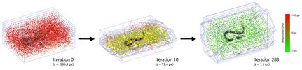
To realize our goal of studying gene expression patterns in the dynamic morphogenetic context, we sought a microscopy technique that would enable high-resolution live imaging throughout the Drosophila embryo. Selective Plane Illumination Microscopy (SPIM) accomplishes in toto imaging of large specimens by acquiring image stacks from multiple angles. We developed an algorithm for registration of multi-angle microscopy acquisitions using fluorescent beads in rigid mounting medium as fiduciary markers (Preibisch et al. 2010). The beads are matched as invariant, local geometric point descriptors and their displacement is minimized for globally optimal, sample-independent registration. We show that the approach can be used for registration of multi-angle acquisition in many imaging modalities.
We recorded Drosophila embryos expressing His-YFP in all cells throughout embryonic development. We are currently exploring the possibilities of segmenting and tracking cells in these datasets. We plan to extend this analysis to record gene expression reporters in the context of the cellular anatomy. SPIM is also ideal for studying cellular level anatomy of fixed specimens in many developmental contexts and will be employed systematically for imaging the FlyFos transgenes.
In order to increase the throughput of SPIM imaging and to make the technology widely available to the developmental biology community, we have initiated an OpenSPIM project to build a custom specialized SPIM set-up on a limited budget. The OpenSPIM will be fully open access both in terms of open hardware and software and the accompanying wiki site will describe in meticulous details all steps necessary for realizing the set-up (OpenSPIM). Thus even laboratories lacking any experience in optical technology development will be able to build such OpenSPIM set-up. We hope that the platform will also serve for further prototyping of more sophisticated SPIMs optimized for various applications.
In collaboration with Tassos Pavlopoulos we are applying the SPIM technology to first describing and next explaining the differential cellular behaviors that lead to morphological and functional differences among appendages on different segments of crustaceans, using Parhyale hawaiensis as a model system.
collaborators: Carl-Zeiss Microimaging Gmbh, Jan Huisken (MPI-CBG), Johannes Schindelin and Kevin Eliceiri (LOCI Madison)
funding: ERC, HFSP 2008
Preibisch, S. W., Saalfeld, S., Schindelin, J., Tomancak, Pavel (2010) Software for bead-based registration of selective plane illumination microscopy data Nature Methods, 7(6), 418-419
Reynaud E., Tomancak P. (2010) The First Light Sheet Based Fluorescence Microscopy Workshop Biotechnology Journal Aug;5(8):798-804
Mohammad S.U., Lee H.K., Preibisch S., Tomancak, P. (2011) Restoring uneven illumination in selective plane illumination microscopy images. Microscopy and Microanalysis Jun 20:1-7