Video reproduced from eLife 2018;7:e34410
During the development of a multicellular organism, a single cell gives rise to a multitude of cell types organized in different tissues and organs. For more than a century now, biologists have been striving to understand the developmental history, referred to as cell lineage, of the different structures in developing organisms. For example, which cells give rise to the eyes or the limbs, and how do cells modify their behaviors to achieve the proper structure and function of the developing organ? Answering such questions has been hindered by difficulties in observing how cells behave in the context of intact developing embryos. To address this limitation, a multinational and multidisciplinary effort was led by the groups of Pavel Tomancak at the Max Planck Institute of Molecular Cell Biology and Genetics (MPI-CBG) and Anastasios Pavlopoulos, former lab member of the Tomancak lab and now located at the HHMI Janelia Research Campus USA. They teamed up with researchers from the Humboldt University and the Max Delbrück Center for Molecular Medicine, both in Berlin, the Pasteur Institute in France, and other colleagues at Janelia to study the cellular basis of limb development.
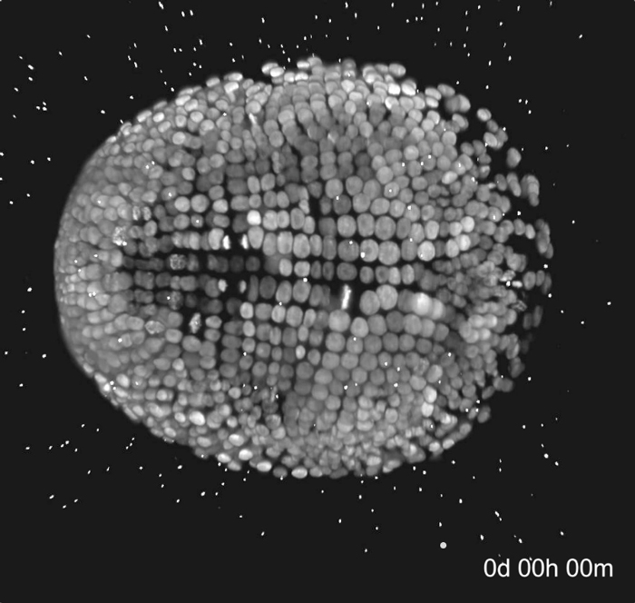
The team focused on the marine amphipod crustacean Parhyale hawaiensis that develops a remarkable suite of specialized limbs along its body axis resembling a living Swiss army knife. Parhyale embryos with fluorescently labeled nuclei were imaged with multi-view light-sheet microscopy. After producing these large multi-dimensional microscopy acquisitions, the researchers faced the challenge how to extract biologically meaningful information from these complex datasets. So, the team developed a user-friendly software, called Massive Multi-view Tracker (MaMuT), freely available as a Fiji/ImageJ plugin. In order to get a comprehensive picture of the developing Parhyale limbs, the embryos had to be imaged and analyzed from multiple angular viewpoints, which was made possible with this combination of multi-view light-sheet microscopy and MaMuT. The researchers produced a very detailed cell lineage of a developing Parhyale limb that was reported in the open-access journal eLife. Anastasios Pavlopoulos explains: “We were finally able to trace and study all cells of growing limbs in intact Parhyale embryos over several days of embryogenesis, from early specification until late differentiation stages”. With these techniques, the researchers could get insights into how cells organize themselves into forming limbs, and could quantify a number of morphogenetic cell behaviors contributing to limb bud formation and elongation.
Pavel Tomancak gives an outlook: “The tools and resources described in this paper will most likely help reveal the cellular and molecular mechanisms that define cell lineages and determine cell fates during the development of an organism. This might help explain the underlying causes of developmental malformations.”
Carsten Wolff et.al: Multi-view light-sheet imaging and tracking with the MaMuT software reveals the cell lineage of a direct developing arthropod limb, eLife, 29 March 2018