Ideally one would like to label and image all structures of interest in a single sample. Up until now filter-based fluorescence imaging prevented us from collecting many colors in parallel: due to the spectral overlap of the fluorophores it has been impossible to distinguish more than four colors.
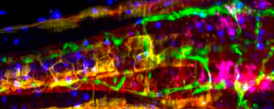
Wiebke Jahr and co-workers in the Huisken lab now made a big step towards imaging many colors in parallel in light sheet microscopy. Instead of acquiring individual colors sequentially with filters, they created a platform based on line-scanning light sheet microscopy to record the entire spectrum for each pixel in a three-dimensional volume. By spectral unmixing they resolved overlapping fluorophores with up to nanometre resolution and removed autofluorescence in zebrafish and fruit fly embryos. The hyperspectral SPIM opens up the possibility to simultaneously image dozens of fluorophores expressed in different tissues of a living organism. A single recording may then provide all the information necessary to study the development and interactions of many tissues that is otherwise difficult to assemble from data sets acquired from several individuals.
Jahr, Wiebke, Benjamin Schmid, Christopher Schmied, Florian O. Fahrbach, and Jan Huisken:
Hyperspectral Light Sheet Microscopy.
Nature Communications 6 (September)
doi:10.1038/ncomms8990.