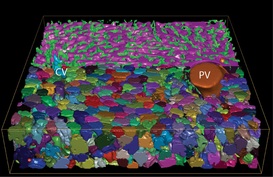
Reconstruction of the main structures forming the liver lobule: Central (CV) and Portal veins (PV), sinusoidal (magenta) and bile canaliculi (green) networks, and hepatocytes (random colours). Copyright: Morales-Navarrete et al. / MPI-CBG
In 1949, Hans Elias pioneered the structural analysis of the mammalian liver tissue and proposed a model of the basic structural unit, the liver lobule, which is used to this day in textbooks. However, this simplified 3D model could only show in a limited way how liver tissue is structured and formed. Almost 70 years later, researchers at the Max Planck Institute of Molecular Cell Biology and Genetics (MPI-CBG), the MPI for the Physics of Complex Systems (MPI-PKS), and the TU Dresden took advantage of novel microscopy developments, computer-aided image analysis, and 3D tissue reconstruction and created a new realistic 3D model of liver organization. Remarkably, they discovered that the liver features an organized structure, similar to liquid crystals. These findings are published in the journal eLife.
The liver is the largest metabolic organ of the human body with a complex tissue architecture. It is vital for blood detoxification and metabolism. Blood flows through blood vessels to the liver cells, called hepatocytes, which take up and metabolize substances and secrete bile for discharge into the intestine. How do cells interact with each other and self-organize to form a functional tissue? For this, its three-dimensional structure must be known. The architecture of tissues and its relation to their function are still poorly understood today. Thus, an interdisciplinary team of biologists, physicists and mathematicians at the MPI-CBG, the MPI-PKS, and the TU Dresden aimed to create a new model of the liver that would be fit to explain how cells collectively form liver tissue and, therefore, a healthy organ.
A structural model of the liver lobule was made and hand-drawn by the anatomist Hans Elias in 1949. Since then, very little progress has been made. To solve this outstanding problem, the Dresden researchers computationally reconstructed the three-dimensional geometry of the tissue from microscopy images of mouse liver tissue and analyzed it applying concepts from Physics. Surprisingly, given the amorphous appearance of liver tissue, the researchers found that the hepatocytes follow a liquid-crystal order, similar to the one making electronic displays. Liquid crystals are less structured than crystals but are more organized than molecules in a liquid. Hernán Morales-Navarrete, postdoctoral researcher in the lab of MPI-CBG director Marino Zerial, explains: “Our results suggest that liver cells and sinusoids, which are the smallest blood vessels in the body, communicate with each other in both directions: The blood vessels instruct the hepatocytes and the hepatocytes send signals back to the blood vessels to establish and preserve the liquid-crystal order. This bi-directional communication is a central part of the self-organization of liver tissue.” Such an architecture gives the tissue function and robustness against local damage.
Marino Zerial, who is also affiliated with the Center for Systems Biology Dresden (CSBD), summarizes: “We discovered novel design principles of liver tissue organization. Only if we understand how the liver tissue is formed to create a functional organ, we will understand abnormalities and malfunctions in humans better. Furthermore, our study provides a general framework, beyond the liver tissue, for clarifying the rules of how cells interact with each other and assemble into tissues.”
Hernán Morales-Navarrete, Hidenori Nonaka, Andre Scholich, Fabián Segovia-Miranda, Walter de Back, Kirstin Meyer, Roman L Bogorad, Victor Koteliansky, Lutz Brusch, Yannis Kalaidzidis, Frank Jülicher, Benjamin M Friedrich, Marino Zerial: “Liquid-crystal organization of liver tissue” eLife, 17. June, 2019. doi: 10.7554/eLife.44860