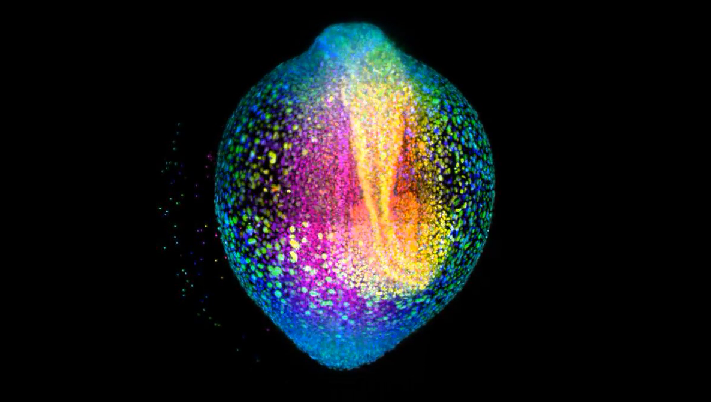
A developing zebrafish embryo
Gopi Shah from the Huisken Lab had participated in a photomicrography competition organized by the camera manufacturing company Andor. The movie acquired on the lab's home-built light sheet microscope setup got first place in the life sciences category for the Andor Insight Award 2015 - congratulations!
The movie shows a developing zebrafish embryo imaged from 4 to 18 hours post fertilization where each cell nucleus is labelled with GFP. Cells are color coded for depth to visualize how dynamic cell reorganization gives rise to the body axis of zebrafish.