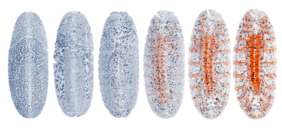
20-h imaging of a fruit fly embryo with the nervous system fluorescently labeled. AutoPilot automatically detects the onset of the expression of the marker and optimizes all parameters associated with this color channel in response to the emerging signal.
Light-sheet microscopy is one of the most powerful methods for imaging the development and function of whole living organisms. However, achieving high-resolution images with these microscopes requires manual adjustments during imaging. Researchers of the Max Planck Institute of Molecular Cell Biology and Genetics in Dresden together with colleagues at Janelia Research Campus (HHMI) have developed a new kind of light-sheet microscope that can ‘drive’ itself automatically by adapting to the challenging and dynamic optical conditions of large living specimens. This new smart microscope combines a novel hardware design and a smart ‘AutoPilot’ system that can analyze images and automatically adjust and optimize the microscope. This framework enables for the first time long-term adaptive imaging of entire developing embryos and improves the resolution of light-sheet microscopes up to five-fold.
The long-standing goal of microscopy is to achieve ever-sharper images deep inside of living samples. In the case of light sheet microscopy, a laser irradiates perpendicularly to the observation direction on the sample to be examined. The laser beam is focused by a lens in only one direction. This creates a light disk illuminating only a thin layer within the sample. In this way, the laser excites dye molecules only in the plane observed by the microscope. Scattered light from other layers, which impairs the image quality, is thus largely avoided.
For light-sheet microscopes this requires to perfectly maintain the careful alignments between imaging and light-sheet illumination planes. Mismatches between these planes arise from the optical variability of living tissues across different locations and over time. Tackling this challenge is essential to acquire the high-resolution images necessary to decipher the biology behind organism development and morphogenesis. “So far, researchers had to sit at their microscope and tweak things manually – our system puts an end to this: it is like a self-driving car - it functions autonomously”, says Loïc Royer, first author of the study. This smart autonomous microscope can in real-time analyze and optimize the spatial relationship between light-sheets and detection planes across the specimen volume.
The researchers demonstrated the performance of their smart microscope by imaging the development of zebrafish and fly embryos for more than 20 hours. They also performed adaptive whole-brain functional imaging in larval zebrafish – obtaining sharper images of a whole ‘thinking’ fish brain. In the study they show how their system recovers cellular and sub-cellular resolution in many regions of the sample and how it adapts to changes in the spatial distribution of fluorescent markers. “We have been using our AutoPilot system on several microscopes for more than two years and it makes a big difference in terms of image quality,” says Philipp Keller, one of the senior authors with Gene Myers. Making microscopes smart and autonomous is important as it will enable the future use of light-sheet microscopy for automated high-throughput drug screens, mutant screens and the construction of anatomical and developmental atlases in various biological model systems.
This work is the result of an international collaboration between the labs of Philipp Keller at Janelia (HHMI, USA) and Gene Myers at MPI-CBG (Dresden, Germany).
Spatiotemporally adaptive imaging of Drosophila embryonic development:
https://vimeo.com/160347546
Spatiotemporally adaptive imaging of dynamic gene expression patterns:
https://vimeo.com/189233797
Spatiotemporally adaptive whole-brain functional imaging in larval zebrafish:
https://vimeo.com/189229181
Loïc A Royer, William C Lemon, Raghav K Chhetri, Yinan Wan, Michael Coleman, Eugene Myers & Philipp J Keller:
Adaptive light-sheet microscopy for long-term, high-resolution imaging in living organisms
Nature Biotechnology, 31 October 2016
doi: 10.1038/nbt.3708