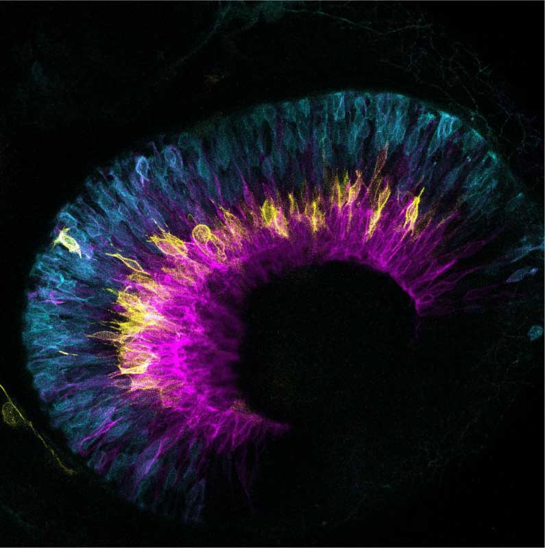
Correct formation of the different neuronal types in the zebrafish retina, here labelled with different colours, is ensured by asymmetric cell divisions. © Nerli et al. / MPI-CBG
Researchers at the at the Instituto Gulbenkian de Ciência (IGC) in Portugal and the Max Planck Institute of Molecular Cell Biology and Genetics (MPI-CBG) in Dresden, Germany have discovered that in the developing retina, and important part of the central nervous system, the divisions leading to the first differentiating neurons are asymmetric. This asymmetry is necessary to generate the correct types of neurons in the right numbers and proportions. The study, published in the scientific journal eLife, is the first to report this asymmetry and the molecular processes underlying it, opening new paths for understanding how the complex brain develops its architecture and function.
Balancing proliferation and differentiation in a developing organ are complex acts, especially when these two processes occur at the same time in the same space. The retina is an important interface between the body and the external world: it sits at the back of our eyes and receives and encodes all the visual information, so that our brain can continuously process the pictures of what the world has to offer. “For attaining this function, the retina requires a precise balance of different types of neurons organised in several interconnected layers, each receiving, pooling or filtering the visual input”, explains Elisa Nerli, first author of the study and researcher at Instituto Gulbenkian de Ciência. “The formation of the different neurons in the correct numbers and proportions is ultimately ensured by balancing cellular proliferation and differentiation during development”.
Studying the development of the zebrafish retina, the team led by Caren Norden, principal investigator at the Instituto Gulbenkian de Ciência and MPI-CBG Alumnus, discovered that this balancing depends on asymmetric divisions of progenitor cells on their way to making functional neurons. “We further found”, Nerli continues, “that the molecular regulation of this process relies on the Notch signalling pathway, since its inhibition interferes with the division asymmetry. We were able to observe that Notch is asymmetrically distributed during cells division. The cell that inherits Notch signalling will continue to proliferate, whereas the other cell will enter a neurogenic lineage.”
This study adds new perspectives to the fundamental understanding of how cellular decisions of proliferation or differentiation can regulate the development of the nervous system. Understanding how the balance between these processes is determined and maintained is important for a better understanding of brain development, in health and in disease.
This study was mainly conducted at the Max Planck Institute of Molecular Cell Biology and Genetics in Dresden, Germany and finished at the IGC. It was funded by the ERC consolidator grant (H2020 ERC-2018-CoG-81904) and the Deutsche Forschungsgemeinschaft (NO 1068/5-1).
Elisa Nerli, Mauricio Rocha-Martins and Caren Norden: Asymmetric neurogenic commitment of retinal progenitors involves Notch through the endocytic pathway. eLife. 03. November 2020. doi: 10.7554/eLife.60462