The skull is essential to human life as it protects the brain from damage. Despite the skull’s importance, many fundamental questions about skull development remain unanswered: What cellular behaviours drive skull growth and morphogenesis during embryogenesis? How is skull expansion regulated genetically? Which cellular processes go wrong in human craniofacial diseases? How does variation between species alter the cellular dynamics of skull growth?
My lab combines a novel ex-vivo live imaging technique, tissue mechanics, and mammalian genetics to understand how the skull grows. We hope to apply our findings to the understanding of human diseases and craniofacial evolution.
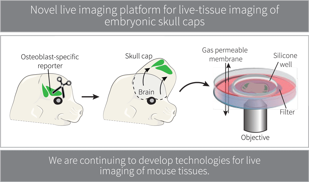
The skull forms a curved bone plate that covers the brain. The curvature of the developing skull and the sheer size of mouse embryos undergoing skull expansion are significant challenges when performing live imaging studies of the developing skull in vivo. We are currently developing new ways to perform in vivo cell imaging of intact craniofacial structures in late stage embryos.
Our goal is to determine how brain growth and size might contribute to the collective cellular dynamics of osteoblasts in the skull. Ultimately, we will test how differences in brain size contribute to the shape and development of the skull between species and the evolution of rapid skull expansion found in mammals.
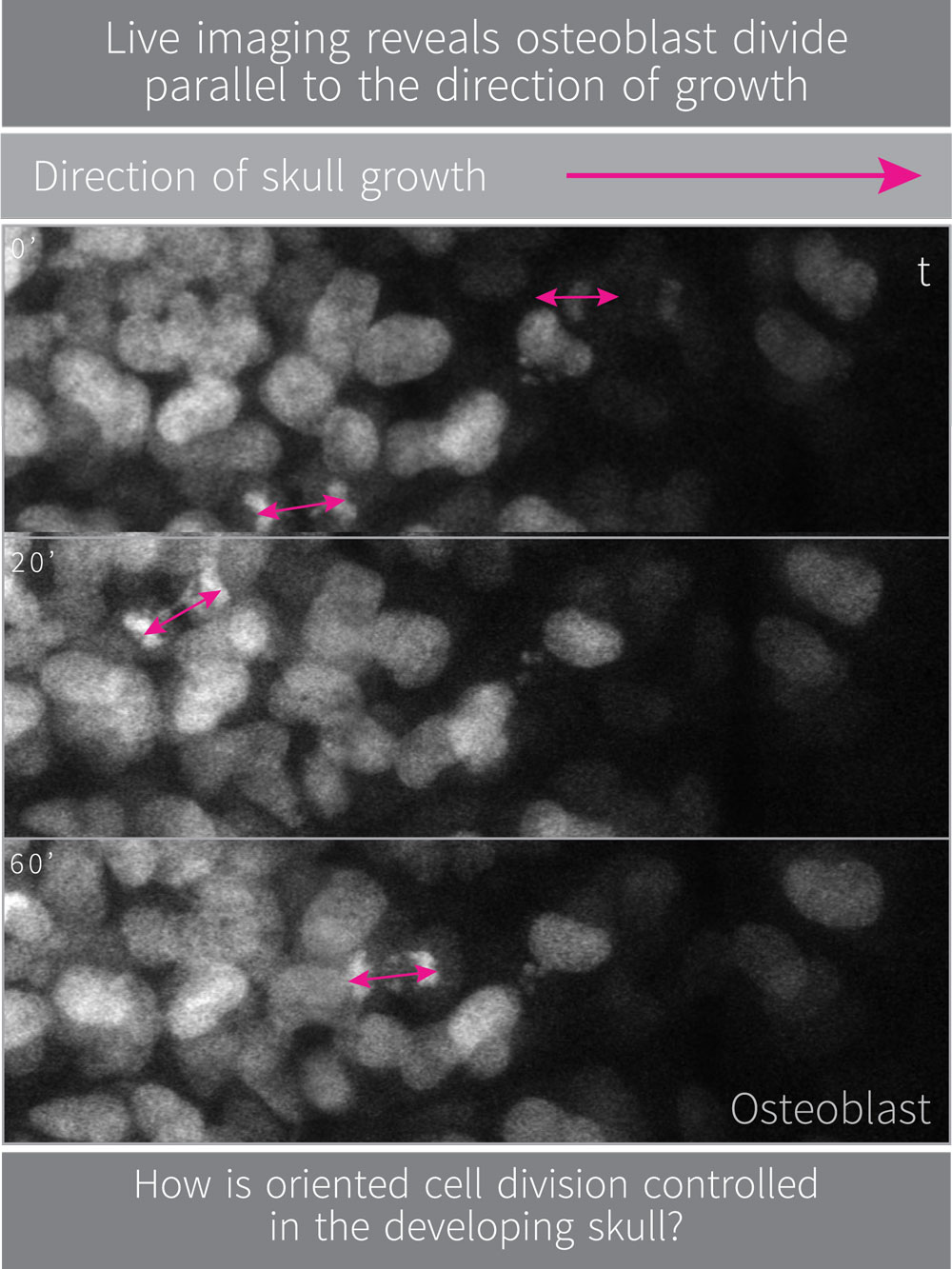
Oriented cell division is used throughout embryonic development and adult homeostasis to shape and pattern tissues. Our live imaging of developing skulls reveals that oriented osteoblast cell division occurs at the growing front of calvaria. The morphogenetic importance and genetic control of oriented cell division in the developing skull is unknown. We study how oriented cell division contributes to skull growth and how this process is regulated genetically.
Osteoblasts also migrate towards the top of the head in addition to undergoing oriented divisions. We study how subcellular dynamics contribute to the expansion of skull bones and migration of osteoblasts.
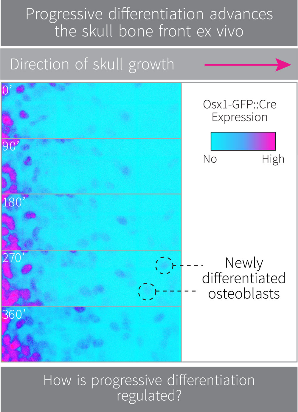
Decades of research have shown that differentiation is the result of stereotypic gene expression changes that are required to specify the fate of a cell. Cell differentiation within a tissue is tightly regulated both in time and space throughout development. Our live imaging studies indicate that newly differentiating osteoblasts are added to the bone front during calvarial expansion, suggesting a progressive wave of oriented differentiation. We are using live imaging, transgenic reporters of osteoblast gene expression and targeted RNAseq approaches to identify the molecular regulators of progressive osteoblast differentiation during expansion of the skull.
Our overall goal is to address fundamental questions in developmental biology and evolution; how are tissues shaped and what are the regulators of tissue shape generation and variation. The mammalian skull is a useful model for tackling these questions as it is a simple sheet-shaped tissue that expands, and like many tissues, does so anistropically. Moreover, skull shape is often altered in craniofacial pathologies and skull development differs significantly between species. By using novel live imaging approaches and mammalian genetics we investigate the dynamic coupling of growth and morphogenesis during skull tissue development.