Former Research Page of Elisabeth Knust, last updated: January 2023
Epithelial tissues in multicellular organisms are characterised by a pronounced apico-basal polarity, which is manifested in the asymmetric distribution of organelles and cytoplasmic proteins, and in the differentiation of the plasma membrane into two distinct domains, the apical and the baso-lateral domains. These domains can be further subdivided into spatially and functionally distinct regions.
Work in our group is aimed to understand the cellular, molecular and genetic mechanisms that control the establishment and maintenance of epithelial cell polarity and that are required to pattern the cell membrane and underlying cytocortex into functionally distinct domains. Studies to answer these questions are performed in the fruitfly Drosophila melanogaster, the zebrafish Danio rerio and in cells in culture.
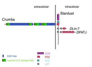
Our research concentrates on the evolutionarily conserved Crumbs (Crb) protein complex. crumbs encodes a large transmembrane protein, with 29 EGF-like repeats and four laminin A G-domain-like repeats in its extracellular domain. The small cytoplasmic domain of only 37 amino acids binds Stardust, a member of the membrane associated guanylate kinase (MAGUK) family. Stardust recruits two additional scaffolding proteins, DLin-7 and DPATJ, into the protein complex.
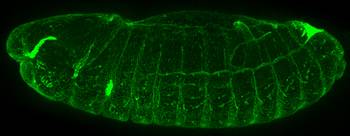
A Drosophila embryo expressing a GFP-tagged Crumbs protein. Anterior is left, dorsal up [Sven Klose, unpublished].
The Crb protein complex is expressed in all epithelia derived from the ectoderm (epidermis, fore- and hindgut, Malpighian tubules, salivary glands), where it is localised in the subapical region apical to the zonula adherens.
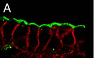
Photoreceptors develop from epithelial cells by a complex morphogenetic process. During this process and in adult photoreceptor cells, the Crumbs protein complex is localised in the stalk membrane, a region of the apical membrane between the rhabdomere and the zonula adherens, which corresponds topologically to the subapical region of epithelial cells. Loss of crumbs, stardust or DPATJ prevents the morphogenesis of the photoreceptor cells.

Cross-section through a Drosophila ommatidum. The apical membrane is subdivided into the most apical part, the rhabdomere (blue), a highly pleated membrane that contains the components of the light-induced signal transduction cascade, and the stalk (red), where the Crb complex is localized [Natalia Bulgakova, unpublished]
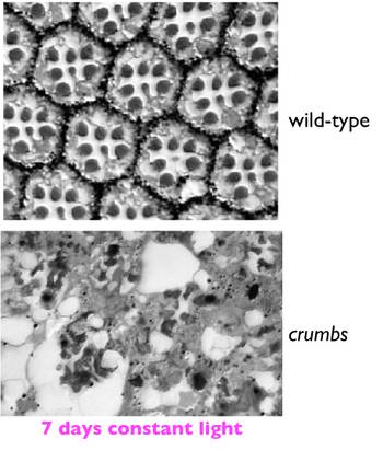
Cross-sections through a wild-type and a crb mutant eye kept in constant illumination for 7 days. Most mutant photoreceptor cells have degenerated [From: Johnson et al., 2002]
crb, sdt and DLin-7 are essential to prevent light-induced retinal degeneration. This phenotype is reminiscent to that induced by mutations in one of the three human crumbs homologues, Crb1, which lead to Retinitis pigmentosa 12, a retinal dystrophy characterised by an early onset blindness. This similarity makes Drosophila an ideal model to study the cellular, molecular and genetic basis of this disease.
Photoreceptors develop from neuroepithelial cells. Their differentiation requires mechanisms to establish and maintain a pronounced apico-basal polarity. As in Drosophila photoreceptor cells, the vertebrate Crb protein complex is localized apical to the adherens junctions, on the inner segment. We are studying the role of members of the Crb complex in the zebrafish retina, in particular during formation and the maintenance of apico-basal polarity.
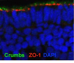
The retina of a zebrafish (72 hours post fertilization). Crb (green) is localized apical to the adherens junction (red). Nuclei are stained in blue [Marta Luz, unpublished].