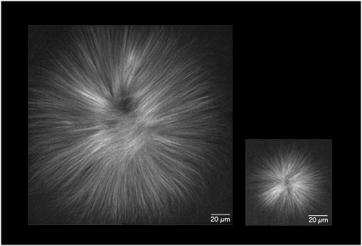
Controlled microtubule creation determines the size of microtubule structures. Picture: MPI-CBG
The size of tissues, cells, and intracellular structures is highly regulated to ensure their proper function. The mitotic spindle, a complex protein machinery in charge of segregating chromosomes during cell division, is a classic example of this regulation. Smaller cells have smaller spindles than bigger cells in the same organism. However, this is not true for very large cells: during the first rounds of cell division after fertilization in an embryo, when cells are very large, spindles reach an upper size limit. How do cells regulate their spindle size? The main building blocks of a spindle are dynamic filaments called microtubules. These filaments only last for about tens of seconds whereas the entire structure can last for hours. Hence, new microtubules have to be constantly created to maintain the structure of the spindle. This process is called microtubule nucleation. However, up to now the underlying mechanisms of microtubule nucleation remained poorly understood.
The research group lead by Jan Brugués at the Max Planck Institute of Molecular Cell Biology and Genetics Dresden and the Max Planck Institute for the Physics of Complex Systems has now developed a method to measure microtubule nucleation events in spindles in isolated cell extracts of the African clawed frog Xenopus laevis. Combining their new method with theoretical analysis and quantitative microscopy, Jan Brugués and his team have shown that the size of a spindle is controlled by a self-amplified growth of microtubules. This growth is regulated by limited amounts of factors only produced at the DNA that induce microtubule nucleation. With these results at hand, the Brugués group has identified a mechanism providing an upper limit to the spindle size in very large cells.
Franziska Decker, the leading author of the recently published paper in eLife, says:
“These findings help us to understand how biological structures are built out of dynamic and short-lived components.” Jan Brugués, who is also member of the neighboring Center for Systems Biology Dresden, adds: “We hope that these findings will help to better understand how cellular structures are set and regulated during embryonic development.”
Franziska Decker, David Oriola, Benjamin Dalton, Jan Brugués: Autocatalytic microtubule nucleation determines the size and mass of Xenopus laevis egg extract spindles, eLife, 11 January 2018