A microsphere is moved with optical tweezers by a well-defined pattern. Movie by Michael Bugiel (BCF)
Optical trap writing
Controlled manipulation of a microsphere for writing. Movie by Michael Bugiel (BCF)
Optical trap pong game
Using a dual optical trap to play 'pong' with two beads. Video by Michael Bugiel (BCF)
Probing a chromosome
A chromosome (labelled with SpyDNA555) is held by two traps and stretched until a single DNA strand is pulled out the chromosome. Overlay of confocal and brightfield microscopy. Movie by Maria Kharlamova (Brugues lab)
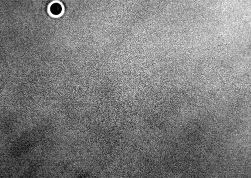
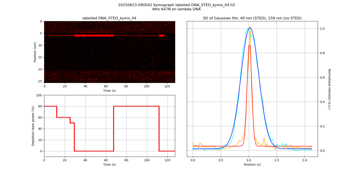
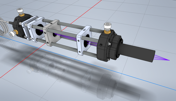
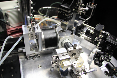
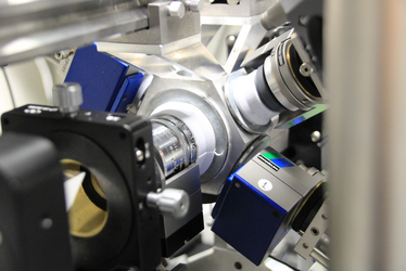
Optical trapping and STED
Super-resolution imaging of a single ATTO 647N dye on DNA, hold by optical tweezers. Image by Michael Bugiel (BCF)
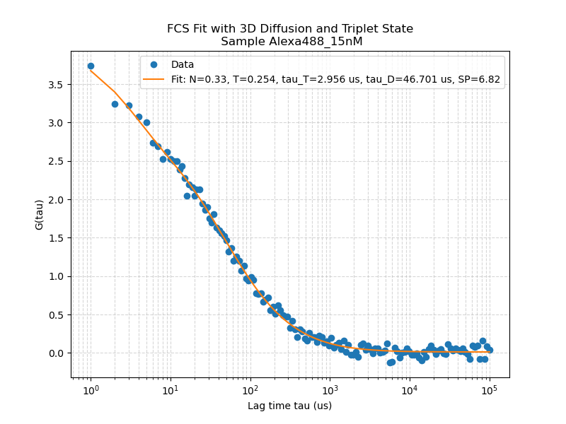
Fluorescence correlation spectroscopy
Autocorrelation function of Alexa-488, gained by FCS in the confocal C-Trap. Sample by Anupam Singh (Zerial lab)
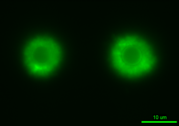
DNA-coated beads
Widefield fluorescence imaging of optically trapped, DNA-coated beads. Image by Franziska Lesche (Brugues group)
Force-dependent labeling of DNA
Widefield fluorescence imaging of DNA which is optically trapped with two beads. At higher loads, more sytox dye binds to the DNA. Movie by Pranay Mandal (Grill group)
Rotation of a microtubule-coated bead
Widefield fluorescent imaging of an optically trapped, microtubule-coated bead that rotates under flow. Movie by Serapion Pyrpassopoulos (Schaeffer group, University of Tuebingen)
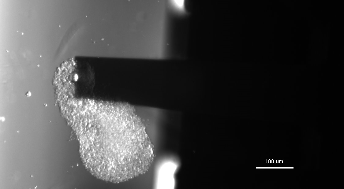
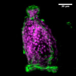
Nanoindentation
Nanoindentation of a gastruloid
Indenting a gastruloid with a 52 um-sized sphere. Image by Marc Trani (Veenvliet group)
Nanoindentation of soft samples
Nanoindenter measurement with indentation, relaxation, and retraction. Sample by Niyousha Baboli (Tabler group)
Nanoindentation of 3D hydrogel structure
Nanoindenter probe (size 52 um) on a PEG-acrylate hydrogel structure. Image by Michael Bugiel (BCF)
Micropatterning
Micropatterning on a fluorescent coverslide
“Writing” the MPI logo by micropatterning on a fluorescent coverslide. Image by Michael Bugiel (BCF)
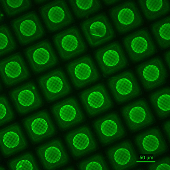
Micropatterning of TEM grids
Selective passivation of PEGylated TEM grids by micropatterning, visualized by GFP binding. Image by Petra Kiesel (von Appen group)
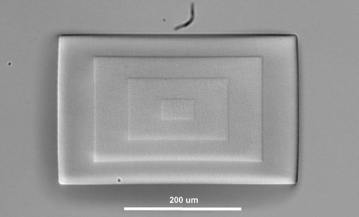
Microstructuration of hydrogels
Hydrogel microstructure as step pyramid, created by photo-polymerization of PEG-acrylate. Image by Michael Bugiel (BCF)
Light sheet microscopy
Pollen grain actin
Structured illumination, sample by Jurai Sekereš
Volvox
XWing scope, movie by Robert Haase (Myers group), sample by Mangal Prakash (Jug group)
C. elegans embryo
Lattice lightsheet, max. projection. Movie by Okafornta William (Müller-Reichert group)
3-D reconstruction of endosomes (green) and mitochondria (red) in a HeLa cell
Lattice lightsheet. Fosheng Hsu (Zerial group)
Zebrafish mitosis
Lattice lightsheet. Maximum projection of an histone-GFP labeled zebrafish embryo.
Drosophila embryo, His-RFP
XWing scope, movie by Robert Haase (Myers group)
C. elegans embryo, His-GFP βTub-GFP
Bessel SPIM, movie by Loïc Royer
Insulin granules
Lattice lightsheet (max projection), 3 volumes/second. Movie by Andreas Müller (Solimena group)
Zebrafish brain acute manipulation. Ras-GFP
Bessel SPIM. Ablation: 920 nm, 500 mW, Ti:Sa laser. Sample by Jaroslav Icha, Imaging by Kei Murata