photo: Emma Miesler
The development of new cutting-edge light microscopy technologies demands the cooperation between many scientists from multiple disciplines. However, after the technology is born, only a few scientists have access to the emerging tool before a commercial version is available for purchasing. A major challenge in using these technologies is therefore a long waiting period for the systems which can be used by a broad community of researchers. For example, the first commercial light sheet microscope, the Z.1, marketed by Zeiss, took 8 years from the original inventor’s publication date to market introduction of the final product. Today it is clear that light sheet microscopy has revolutionized the way we visualize and make sense of complex biological processes. This example shows that faster access to emerging technologies is crucial for the advancement of science. To shorten the gap between invention of an imaging technology and its regular use in research, the newly stablished Advanced Imaging Facility, located at the MPI-CBG and the Center for Systems Biology Dresden, offers a unique opportunity for developers and users to build, test and improve novel light microscopy technologies before they become commercially available.
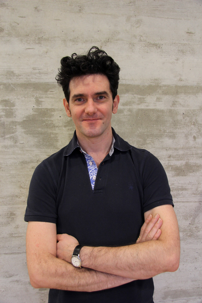
Nicola Maghelli, Service Leader of the AIF provides the inside story:
How is AIF different from the outstanding Light Microscopy Facility (LMF) at the MPI-CBG?
AIF, does not stand for “Another Imaging Facility”. Actually, these two facilities complement each other in supporting scientists and accelerating scientific discoveries. Also, users should not just come to me because the “A” in the facility’s name. I strongly recommend users to first check with the LMF team if they can support their imaging needs.
The LMF provides users with a vast arrange of state-of-the-art commercial instruments that are serviced by the manufactures. The AIF on the other hand, builds and maintains advanced light microscopy setups that you simply cannot buy in the market today. Because of our expertise in the fields of optics, opto-mechanics and instrument control, we actively cooperate with research groups who are developing their own custom setups. These technologies can be duplicated and further developed at the facility, making it accessible to early adopters.
How does AIF support the scientific community?
We support our users in three different ways. Firstly, we help individual users to choose the most suited system for their samples and we assist them during the whole imaging workflow. Additionally, we can help with post-processing the raw data. Secondly, the facility can support research groups who want to develop their own custom setups. We can provide them with the technical know-how and expertise. We can help with the design of optical systems. These systems can then be put together by integration of our custom designed optical and mechanical components. Thirdly, we aim to be a hub for the community of instrument developers. By integrating their ideas, the equipment and the collective expertise, we hope to accelerate the development of novel instruments.
What areas does the AIF specialise in?
The facility houses a number of custom light sheet setups including a lattice light sheet microscope (LLSM). This microscope achieves high precision of optical sectioning and is best suited for thin, transparent specimens. Currently, it is the most popular setup in the facility and its users like the speed, resolution and low phototoxicity of the microscope. Features include structured illumination and GPU-accelerated deconvolution.
Moreover, we are working together with the MPI-CBG research group of Alf Honigmann on building a light sheet microscope for organoids imaging. Our custom microscope is based on a multiview SPIM which was originally developed by the former MPI-CBG research group leader Jan Huisken. We completely redesigned the sample chamber, adding temperature and CO2 control, a perfusion system as well as a magnetic stirrer to keep the imaging medium as homogeneous as possible during an experiment. We aim at imaging samples over multiple days. This setup complements two Zeiss Light Sheet Z.1. microscopes hosted at the LMF which are also suited for organoids imaging. Finally, there are plans for incorporating the “X-wing” microscope in the facility. This light sheet microscope uses four illumination objectives and two high-NA objectives on the detection sides. However, this won’t happen immediately, as the setup is rather complex and still under development in the Myers group.
What is the outlook for the AIF?
Designing the AIF was partly inspired by the Advanced Imaging Center at the Janelia Research Campus in the USA. This center provides cutting edge pre-commercial microscopes, developed at Janelia, available to visiting scientists. In Germany, our AIF is the first facility of its kind. Dresden is a vibrant hub for experts in biology, optics, imaging, computer science and bio-image informatics. Consequently, the facility is an ideal place for developing novel solutions and technologies while making revolutionary non-commercial technologies available for biologists ahead of their time.
In the broader context, supposing that there will be more funds available, there are bigger plans to transform our facility into cAID – Center of Advanced Imaging Dresden. The idea is to create a very unique place in Germany (and Europe, maybe!) for advanced imaging. I really do hope that this will happen!