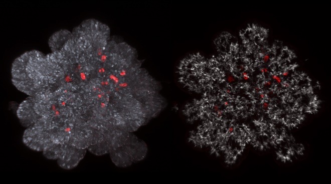
Artificial three-dimensional niches deconstruct pancreas development in vitro
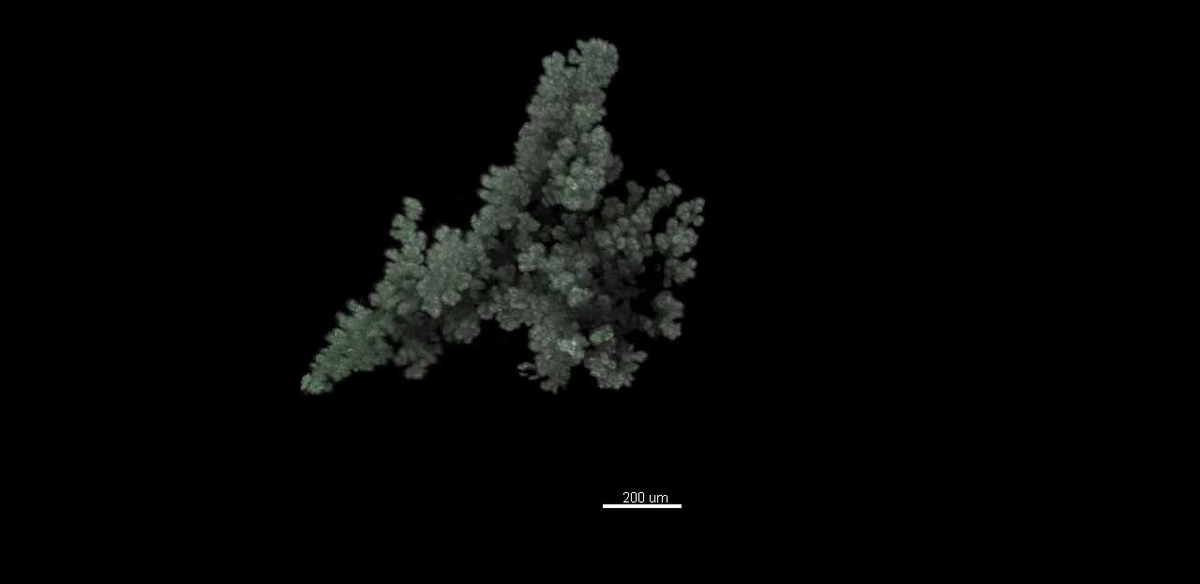
We developed the first pancreas organoid model, starting from embryonic mouse pancreas progenitors. We showed that cell cooperation is needed to form organoids.
Using genetic lineage tracing, we discovered that the potential of individual pancreatic cells during development is heterogeneous at a given time point of development. We further showed the clonal and complementary live imaging data to be best modelled by a stochastic decision making process.
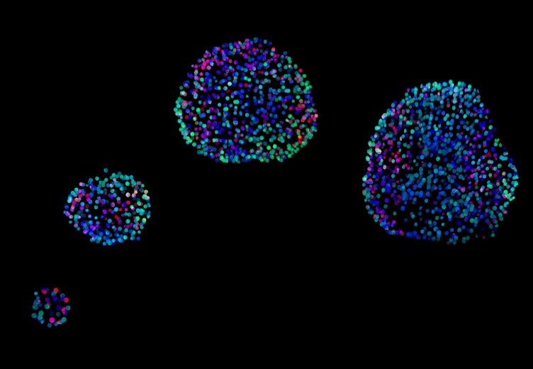
A 3D system to model human pancreas development and its reference single cell transcriptome atlas reveal signaling pathways required for progenitor expansion
We established an organoid model of human pancreas development that we compared to human fetal pancreas by single cell sequencing. We showed its power in screening, identifying molecules that promote pancreas progenitor proliferation.
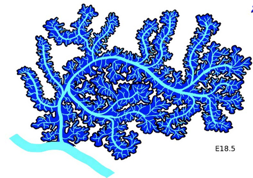
Deconstructing the principles of ductal network formation in the pancreas
We digitized the network of ducts of the pancreas and analysed its structure. Our results showed that its network properties change during development from a mesh to a tree. We proposed that flow starts during development and drives the remodelling. We recently found that pancreatic cells secrete pancreatic juice from very early during development which may create a flow controlling the organization of the ductal tree and possibly differentiation.
Julie Warin, Anne Grapin-Botton Rethinking HNF1A-MODY: HNF1A at the crossroads of development and multiorgan metabolic disease. Genes Dev, 40(11-12) 777-779 (2026)
Open Access DOI
In this issue of Genes & Development, Unger and colleagues (doi:10.1101/gad.353153.125) combined human pluripotent stem cell-derived in vitro models with targeted in vivo mouse models to reveal multiple developmental defects triggered by HNF1A mutations causing maturity-onset diabetes of the young. This work paints the picture of a disorder that starts well before diabetes manifests, highlighting its complexity arising from the diverse roles of HNF1A across distinct cell types, each potentially differentially impacted by different mutations.
Vaibhav Mahajan, Keshav Gajendra Babu, Markus Mukenhirn, Antje Garside, Vinita Ajit Kini, Trishla Adhikari, Timon Beck, Byung Ho Lee, Kyoohyun Kim, Carsten Werner, Raimund Schlüßler, Alf Honigmann, Sebastian Aland, Anna Verena Taubenberger Cells Dynamically Adapt Their Nuclear Volumes and Proliferation Rates During Single to Multicellular Transitions. Adv Sci (Weinh), 13(31) Art. No. e24325 (2026)
Open Access DOI
Tumor development and progression involve biophysical changes across spatial scales, from the subcellular to the multicellular tissue scale. While cells are known to dynamically regulate their volumes and mechanics in dependence of cell state and function, it is unclear how these properties are controlled in dense multicellular environments like developing tumors. Here, we quantified cell and nuclear volumes of cancer cells forming multicellular spheroids within mechanically tunable biohybrid polymer hydrogels. We quantitatively showed that formation of multicellular structures is associated with marked reductions of cellular and nuclear volumes, cell cycle delays as well as cell mechanical alterations, and that these changes are coupled. Single-to-multicellular transitions led to up to 60% decreases in median nuclear volumes, which was not explained by growth-induced compressive stress. Instead, nuclear volume reductions in emerging clusters arose from cell cycle adaptations, with accumulation of smaller G1-phase cells-reversed by CDK1 inhibition. Additional nuclear downsizing in forming clusters was associated with cell mass density and stiffness increases and reverted upon cell release. Conversely, multicellular-to-single cell transitions during invasion were accompanied by nuclear volume expansion and cell softening. Together, these findings reveal dynamic regulation of cellular and nuclear volumes, mechanics, and cell cycle progression in response to multicellular state.
Jifeng Liu, Anne Grapin-Botton Benchmarking in vivo and in vitro gene co-expression networks enables efficient β-like cell differentiation. Dev Cell, 61(2) 229-231 (2026)
DOI
In this issue of Developmental Cell, Yu et al.1 reconstruct human embryonic gene co-expression networks (GCNs) that guide pancreatic endocrine lineage specification. Benchmarking stem cell-derived islet (SC-islet) differentiation protocols against fetal human pancreatic cells reveals early regulatory divergences, thereby enabling the design of a protocol with improved β-like cell production.
Rashmiparvathi Keshara, Karolina Kuodyte, Antje Janosch, Cordula Andree, Marc Bickle, Martin Stöter, Rico Barsacchi, Yung Hae Kim, Anne Grapin-Botton High-content screening of organoids reveals the mechanisms of human pancreas acinar specification. Cell Stem Cell, 33(2) 325-339 (2026)
Open Access DOI
Organoids derived from pluripotent stem cells have emerged as powerful models to study human development. To investigate signaling pathways regulating human pancreas differentiation and morphogenesis, we developed a high-content, image-based screen and quantitative multivariate analysis pipelines robust to heterogeneity to extract single-cell and organoid features using pancreatic progenitor organoids. Here, we identified 54 compounds affecting cell identity and/or morphological landscape. Focusing on one family of compounds, we found that glycogen synthase kinase 3α/β (GSK3A/B) inhibition via wingless/int-1 (WNT) signaling has a reversible effect on cell identity, repressing pancreatic progenitor markers and inducing a poised state in progenitors transitioning to acinar cells. We show that additional fibroblast growth factor (FGF) repression enables further differentiation of acinar cells, recapitulating pancreatic acinar morphogenesis and function. The ability to produce acinar cells is valuable for future studies on pancreatic exocrine function and cancer initiation in humans, as acinar cells are thought to be an important cell of origin for pancreatic adenocarcinoma.
Byung Ho Lee#, Kana Fuji, Heike Petzold, Philip Allan Seymour, Siham Yennek, Coline Schewin, Allison Lewis, Daniel Riveline, Tetsuya Hiraiwa, Masaki Sano, Anne Grapin-Botton# Permeability-driven pressure and cell proliferation control lumen morphogenesis in pancreatic organoids. Nat Cell Biol, 28(1) 113-124 (2026)
Open Access DOI
Lumen formation in organ epithelia involves processes such as polarization, secretion, exocytosis and contractility, but what controls lumen shape remains unclear. Here we study how lumina develop spherical or complex structures using pancreatic organoids. Combining computational phase-field modelling and experiments, we found that lumen morphology depends on the balance between cell cycle duration and lumen pressure, low pressure and high proliferation produce complex shapes. Manipulating proliferation and lumen pressure can alter or reverse lumen development both in silico and in vitro. Increasing epithelial permeability reduces lumen pressure, converting from spherical to complex lumina. During pancreas development, the epithelium is initially permeable and becomes sealed, experimentally increasing permeability at late stages impairs ductal morphogenesis. Overall, our work underscores how proliferation, pressure and permeability orchestrate lumen shape, offering insights for tissue engineering and cystic disease treatment.
2025
Sakurako Tanida, Kana Fuji, Linjie Lu, Tristan Guyomar, Byung Ho Lee, Alf Honigmann, Anne Grapin-Botton, Daniel Riveline, Tetsuya Hiraiwa#, Makiko Nonomura#, Masaki Sano# Predicting organoid morphology through a phase field model: Insights into cell division and lumenal pressure. PLoS Comput Biol, 21(8) Art. No. e1012090 (2025)
Open Access DOI
Organoids are ideal systems to predict the phenotypes of organs. However, there is currently a lack of understanding regarding the generalized rules that enable use of simple cellular principles to make morphological predictions of entire organoids. Therefore, we employed a phase field model with the following basic components: the minimum conditions for the timing and volume of cell division, lumen nucleation rules, and lumenal pressure. Through our model, we could compute and generate a myriad of organoid phenotypes observed till date. We propose morphological indices necessary to characterize the shapes and construct phase diagrams and show their dependencies on proliferation time and lumen pressure. Additionally, we introduced the lumen-index parameter, which helped in examining the criteria to maintain organoids as spherical structures comprising a single layer of cells and enclosing an intact lumen. Finally, we predict a star-like organoid phenotype that did not undergo differentiation, suggesting that the volume constraint during cell division may determine the final phenotype. In summary, our approach provides researchers with guidelines to test the mechanisms of self-organization and predict the shape of organoid.
Rimvile Prokarenkaite, Karolina Kuodyte, Greta Gudoityte, Elzbieta Budginaite, Daniel Naumovas, Egle Strainiene, Kristijonas Velickevicius, Audrius Dulskas, Ernestas Sileika, Jonas Venius, Virginijus Tunaitis, Augustas Pivoriunas, Vytaute Starkuviene, Vaidotas Stankevicius#, Kestutis Suziedelis# PARP9-PARP13-PARP14 axis tunes colorectal cancer response to radiotherapy. J Exp Clin Cancer Res, 44(1) Art. No. 199 (2025)
Open Access DOI
Colorectal cancer (CRC) is the third most prevalent cancer worldwide. Despite substantial advancements in CRC therapy in recent years, ionizing radiation (IR) continues to be the predominant treatment for colon malignances. However, it still lacks the precision required for excellent therapeutic outcomes, ultimately resulting in tumor radioresistance. This study seeks to explore the potential of atypical PARPs including PARP9, PARP12, PARP13 and PARP14 as innovative radiosensitizing targets for CRC.
Linjie Lu*, Kana Fuji*, Tristan Guyomar, Michèle Lieb, Marie André, Sakurako Tanida, Makiko Nonomura, Tetsuya Hiraiwa, Yara Alcheikh, Siham Yennek, Heike Petzold, Cécilie Martin-Lemaitre, Anne Grapin-Botton#, Alf Honigmann#, Masaki Sano#, Daniel Riveline# Generic comparison of lumen nucleation and fusion in epithelial organoids with and without hydrostatic pressure. Nat Commun, 16(1) Art. No. 6307 (2025)
Open Access DOI
Many internal organs in the body harbor a fluid-filled lumen. Lumen nucleation and fusion have been reported as dependent on organ-type during organogenesis. In contrast, the physics of lumen suggests that force balance between luminal pressure and cell mechanics leads to generic rules. However, this hypothesis lacks experimental evidence. Here we compare lumen dynamics for three different systems (MDCK cysts, pancreatic spheres, and epiblast model) by using quantitative cell biology, microfabrication, and theory. We report that the initial cell number determines the maximum number of lumens but does not impact the steady state, which is a final single lumen. We show that lumen dynamics is determined by luminal hydrostatic pressure. We also use MDCK cysts to manipulate cell adhesion and lumen volume to successfully reproduce the fusion dynamics of pancreatic spheres and epiblasts. Our results reveal self-organisation rules of lumens with relevance for morphogenesis and tissue engineering.
Anne Grapin-Botton#, Jonathan Y-H Loh# Editorial overview: Regaining architecture and cell cross-talk upon regeneration. Curr Opin Genet Dev, 91 Art. No. 102302 (2025)
DOI
2024
Tzer Han Tan, Aboutaleb Amiri, Irene Seijo-Barandiaran, Michael F Staddon, Anne Materne, Sandra Tomas, Charlie Duclut, Marko Popović#, Anne Grapin-Botton#, Frank Jülicher# Emergent chirality in active solid rotation of pancreas spheres. PRX Life, 2(3) Art. No. 033006 (2024)
Open Access DOI
Collective cell dynamics play a crucial role in many developmental and physiological contexts. While two-dimensional (2D) cell migration has been widely studied, how three-dimensional (3D) geometry and topology interplay with collective cell behavior to determine dynamics and functions remains an open question. In this work, we elucidate the biophysical mechanism underlying rotation in spherical tissues, a phenomenon widely reported both in vivo and in vitro. Using murine pancreas-derived organoids as a model system, we find that epithelial spheres exhibit persistent rotation, rotational axis drift, and rotation arrest. Using a 3D vertex model, we demonstrate how the combined action of traction force and polarity alignment can account for these distinct rotational dynamics near a solid to flow transition. Furthermore, our analysis shows that the spherical tissue rotates as an active solid occasionally switching to a flowing state and exhibits spontaneous chiral symmetry breaking. Using a continuum model, we demonstrate how the topological defects in the polarity field underlie this symmetry breaking process, which is revealed by asymmetries in the cell elongation pattern. For cell elongation to reveal the chiral asymmetry, shear flow is required in addition to the solid body rotation. Altogether, our work reveals a robust chiral symmetry breaking mechanism with potential implications for left-right symmetry breaking processes in morphogenetic events.
2023
Belin Selcen Beydag-Tasöz, Joyson Verner D'Costa, Lena Hersemann, Byung Ho Lee, Federica Luppino, Yung Hae Kim, Christoph Zechner, Anne Grapin-Botton Integrating single-cell imaging and RNA sequencing datasets links differentiation and morphogenetic dynamics of human pancreatic endocrine progenitors. Dev Cell, 58(21) 2292-2308 (2023)
Open Access DOI
Basic helix-loop-helix genes, particularly proneural genes, are well-described triggers of cell differentiation, yet information on their dynamics is limited, notably in human development. Here, we focus on Neurogenin 3 (NEUROG3), which is crucial for pancreatic endocrine lineage initiation. By monitoring both NEUROG3 gene expression and protein in single cells using a knockin dual reporter in 2D and 3D models of human pancreas development, we show an approximately 2-fold slower expression of human NEUROG3 than that of the mouse. We observe heterogeneous peak levels of NEUROG3 expression and reveal through long-term live imaging that both low and high NEUROG3 peak levels can trigger differentiation into hormone-expressing cells. Based on fluorescence intensity, we statistically integrate single-cell transcriptome with dynamic behaviors of live cells and propose a data-mapping methodology applicable to other contexts. Using this methodology, we identify a role for KLK12 in motility at the onset of NEUROG3 expression.
Jay Gopalakrishnan#, Kerstin Feistel, Benjamin Friedrich, Anne Grapin-Botton, Nathalie Jurisch-Yaksi, Elvira Mass, David U Mick, Roman-Ulrich Müller, Helen May-Simera, Bernhard Schermer, Miriam Schmidts, Peter Walentek, Dagmar Wachten# Emerging principles of primary cilia dynamics in controlling tissue organization and function. EMBO J, 42(21) Art. No. e113891 (2023)
Open Access DOI
Primary cilia project from the surface of most vertebrate cells and are key in sensing extracellular signals and locally transducing this information into a cellular response. Recent findings show that primary cilia are not merely static organelles with a distinct lipid and protein composition. Instead, the function of primary cilia relies on the dynamic composition of molecules within the cilium, the context-dependent sensing and processing of extracellular stimuli, and cycles of assembly and disassembly in a cell- and tissue-specific manner. Thereby, primary cilia dynamically integrate different cellular inputs and control cell fate and function during tissue development. Here, we review the recently emerging concept of primary cilia dynamics in tissue development, organization, remodeling, and function.
Tzer Han Tan, Jifeng Liu, Anne Grapin-Botton Mapping and exploring the organoid state space using synthetic biology. Semin Cell Dev Biol, 141 23-32 (2023)
DOI
The functional relevance of an organoid is dependent on the differentiation, morphology, cell arrangement and biophysical properties, which collectively define the state of an organoid. For an organoid culture, an individual organoid or the cells that compose it, these state variables can be characterised, most easily by transcriptomics and by high-content image analysis. Their states can be compared to their in vivo counterparts. Current evidence suggests that organoids explore a wider state space than organs in vivo due to the lack of niche signalling and the variability of boundary conditions in vitro. Using data-driven state inference and in silico modelling, phase diagrams can be constructed to systematically sort organoids along biochemical or biophysical axes. These phase diagrams allow us to identify control strategies to modulate organoid state. To do so, the biochemical and biophysical environment, as well as the cells that seed organoids, can be manipulated.
Belin Selcen Beydag-Tasöz, Siham Yennek, Anne Grapin-Botton Towards a better understanding of diabetes mellitus using organoid models. Nat Rev Endocrinol, 19(4) 232-248 (2023)
DOI
Our understanding of diabetes mellitus has benefited from a combination of clinical investigations and work in model organisms and cell lines. Organoid models for a wide range of tissues are emerging as an additional tool enabling the study of diabetes mellitus. The applications for organoid models include studying human pancreatic cell development, pancreatic physiology, the response of target organs to pancreatic hormones and how glucose toxicity can affect tissues such as the blood vessels, retina, kidney and nerves. Organoids can be derived from human tissue cells or pluripotent stem cells and enable the production of human cell assemblies mimicking human organs. Many organ mimics relevant to diabetes mellitus are already available, but only a few relevant studies have been performed. We discuss the models that have been developed for the pancreas, liver, kidney, nerves and vasculature, how they complement other models, and their limitations. In addition, as diabetes mellitus is a multi-organ disease, we highlight how a merger between the organoid and bioengineering fields will provide integrative models.
2022
Sean P A Ritter, Logan A Brand, Shelby L Vincent, Albert Remus R Rosana, Allison Lewis, Denise S Whitford, George W Owttrim Multiple Light-Dark Signals Regulate Expression of the DEAD-Box RNA Helicase CrhR in Synechocystis PCC 6803. Cells, 11(21) Art. No. 3397 (2022)
Open Access DOI
Since oxygenic photosynthesis evolved in the common ancestor of cyanobacteria during the Archean, a range of sensing and response strategies evolved to allow efficient acclimation to the fluctuating light conditions experienced in the diverse environments they inhabit. However, how these regulatory mechanisms are assimilated at the molecular level to coordinate individual gene expression is still being elucidated. Here, we demonstrate that integration of a series of three distinct light signals generate an unexpectedly complex network regulating expression of the sole DEAD-box RNA helicase, CrhR, encoded in Synechocystis sp. PCC 6803. The mechanisms function at the transcriptional, translational and post-translation levels, fine-tuning CrhR abundance to permit rapid acclimation to fluctuating light and temperature regimes. CrhR abundance is enhanced 15-fold by low temperature stress. We initially confirmed that the primary mechanism controlling crhR transcript accumulation at 20 °C requires a light quantity-driven reduction of the redox poise in the vicinity of the plastoquinone pool. Once transcribed, a specific light quality cue, a red light signal, was required for crhR translation, far-red reversal of which indicates a phytochrome-mediated mechanism. Examination of CrhR repression at 30 °C revealed that a redox- and light quality-independent light signal was required to initiate CrhR degradation. The crucial role of light was further revealed by the observation that dark conditions superseded the light signals required to initiate each of these regulatory processes. The findings reveal an unexpected complexity of light-dark sensing and signaling that regulate expression of an individual gene in cyanobacteria, an integrated mechanism of environmental perception not previously reported.
Keiichi Katsumoto#, Siham Yennek, Chunguang Chen, Luis Fernando Delgadillo Silva, Sofia Traikov, Dror Sever, Ajuna Azad, Jingdong Shan, Seppo Vainio, Nikolay Ninov, Stephan Speier, Anne Grapin-Botton# Wnt4 is heterogeneously activated in maturing β-cells to control calcium signaling, metabolism and function. Nat Commun, 13(1) Art. No. 6255 (2022)
Open Access DOI
Diabetes is a multifactorial disorder characterized by loss or dysfunction of pancreatic β-cells. β-cells are heterogeneous, exhibiting different glucose sensing, insulin secretion and gene expression. They communicate with other endocrine cell types via paracrine signals and between β-cells via gap junctions. Here, we identify the importance of signaling between β-cells via the extracellular signal WNT4. We show heterogeneity in Wnt4 expression, most strikingly in the postnatal maturation period, Wnt4-positive cells, being more mature while Wnt4-negative cells are more proliferative. Knock-out in adult β-cells shows that WNT4 controls the activation of calcium signaling in response to a glucose challenge, as well as metabolic pathways converging to lower ATP/ADP ratios, thereby reducing insulin secretion. These results reveal that paracrine signaling between β-cells is important in addition to gap junctions in controling insulin secretion. Together with previous reports of WNT4 up-regulation in obesity our observations suggest an adaptive insulin response coordinating β-cells.
Anne Grapin-Botton, Yung Hae Kim Pancreas organoid models of development and regeneration. Development, 149(20) Art. No. dev201004 (2022)
DOI
Organoids have become one of the fastest progressing and applied models in biological and medical research, and various organoids have now been developed for most of the organs of the body. Here, we review the methods developed to generate pancreas organoids in vitro from embryonic, fetal and adult cells, as well as pluripotent stem cells. We discuss how these systems have been used to learn new aspects of pancreas development, regeneration and disease, as well as their limitations and potential for future discoveries.
Rashmiparvathi Keshara#, Yung Hae Kim#, Anne Grapin-Botton# Organoid Imaging: Seeing Development and Function. Annu Rev Cell Dev Biol, 38 447-466 (2022)
DOI
Organoids are miniaturized and simplified versions of an organ produced in vitro from stem or progenitor cells. They are used as a model system consisting of multiple cell types forming an architecture relevant to the organ and carrying out the function of the organ. They are a useful tool to study development, homeostasis, regeneration, and disease. The imaging of organoids has become a pivotal method to visualize and understand their self-organization, symmetry breaking, growth, differentiation, and function. In this review, we discuss imaging methods, how to analyze these images, and challenges in organoid research.
Akiko Nakamura*, Yan Fung Wong*, Andrea Venturato, Magali Michaut, Seshasailam Venkateswaran, Mithun Santra, Carla A C Gonçalves, Michael Larsen, Marit Leuschner, Yung Hae Kim, Joshua Brickman, Mark Bradley, Anne Grapin-Botton Long-term feeder-free culture of human pancreatic progenitors on fibronectin or matrix-free polymer potentiates β cell differentiation. Stem Cell Rep, 17(5) 1215-1228 (2022)
Open Access DOI
With the aim of producing β cells for replacement therapies to treat diabetes, several protocols have been developed to differentiate human pluripotent stem cells to β cells via pancreatic progenitors. While in vivo pancreatic progenitors expand throughout development, the in vitro protocols have been designed to make these cells progress as fast as possible to β cells. Here, we report on a protocol enabling a long-term expansion of human pancreatic progenitors in a defined medium on fibronectin, in the absence of feeder layers. Moreover, through a screening of a polymer library we identify a polymer that can replace fibronectin. Our experiments, comparing expanded progenitors to directly differentiated progenitors, show that the expanded progenitors differentiate more efficiently into glucose-responsive β cells and produce fewer glucagon-expressing cells. The ability to expand progenitors under defined conditions and cryopreserve them will provide flexibility in research and therapeutic production.
Anne Grapin-Botton#, Barbara Ludwig# Stem cell-derived β cells go in monkeys. Cell Stem Cell, 29(4) 500-502 (2022)
DOI
Du et al. transplanted β cells derived from pluripotent stem cells in diabetic monkeys for the first time, as an intermediate stage toward clinical translation. They observed benefits unfolding over months but also observed immune rejection of the grafts by 5-6 months.
Byung Ho Lee*, Irene Seijo-Barandiaran*, Anne Grapin-Botton Epithelial morphogenesis in organoids. Curr Opin Genet Dev, 72 30-37 (2022)
DOI
Epithelial organoids can recapitulate many processes reminiscent of morphogenesis in vivo including lumen and multilayer formation, folding, branching, delamination and elongation. While being noisier in vitro than in vivo, these processes can be monitored live and subjected to interferences, a field that is emerging. We elaborate on the signalling molecules controlling morphogenesis, from the medium and their emergence as signalling centers in the organoids. Further, we discuss how organoid shape is controlled by mechanical cues within the organoid and their interplay with the material properties of the environment.
Zahra Ghezelayagh*, Mahsa Zabihi*, Ibrahim Zarkesh, Carla A C Gonçalves, Michael Larsen, Newsha Hagh-Parast, Mohammad Pakzad, Massoud Vosough, Babak Arjmand, Hossein Baharvand, Banafshé Larijani, Anne Grapin-Botton, Hamid Reza Aghayan#, Yaser Tahamtani# Improved Differentiation of hESC-Derived Pancreatic Progenitors by Using Human Fetal Pancreatic Mesenchymal Cells in a Micro-scalable Three-Dimensional Co-culture System. Stem Cell Rev Rep, 18(1) 360-377 (2022)
DOI
Mesenchymal cells of diverse origins differ in gene and protein expression besides producing varying effects on their organ-matched epithelial cells' maintenance and differentiation capacity. Co-culture with rodent's tissue-specific pancreatic mesenchyme accelerates proliferation, self-renewal, and differentiation of pancreatic epithelial progenitors. Therefore, in our study, the impact of three-dimensional (3D) co-culture of human fetal pancreatic-derived mesenchymal cells (hFP-MCs) with human embryonic stem cell-derived pancreatic progenitors (hESC-PPs) development towards endocrine and beta cells was assessed. Besides, the ability to maintain scalable cultures combining hFP-MCs and hESC-PPs was investigated. hFP-MCs expressed many markers in common with bone marrow-derived mesenchymal stem cells (BM-MSCs). However, they showed higher expression of DESMIN compared to BM-MSCs. After co-culture of hESC-PPs with hFP-MCs, the pancreatic progenitor (PP) spheroids generated in Matrigel had higher expression of NGN3 and INSULIN than BM-MSCs co-culture group, which shows an inductive impact of pancreatic mesenchyme on hESC-PPs beta-cells maturation. Pancreatic aggregates generated by forced aggregation through scalable AggreWell system showed similar features compared to the spheroids. These aggregates, a combination of hFP-MCs and hESC-PPs, can be applied as an appropriate tool for assessing endocrine-niche interactions and developmental processes by mimicking the pancreatic tissue.
2021
Belin Selcen Beydag-Tasöz, Joyson Verner D'Costa, Lena Hersemann, Federica Luppino, Yung Hae Kim, Christoph Zechner, Anne Grapin-Botton A combined transcriptional and dynamic roadmap of single human pancreatic endocrine progenitors reveals proliferative capacity and differentiation continuum. bioRxiv, Art. No. https://doi.org/10.1101/2021.12.15.472220 (2021)
Open Access DOI
Basic helix-loop-helix genes, particularly proneural genes, are well-described triggers of cell differentiation, yet limited information exists on their dynamics, notably in human development. Here, we focus on Neurogenin 3 (NEUROG3), which is crucial for pancreatic endocrine lineage initiation. Using a double reporter to monitor endogenous NEUROG3 transcription and protein expression in single cells in 2D and 3D models of human pancreas development, we show peaks of expression for the RNA and protein at 22 and 11 hours respectively, approximately two-fold slower than in mice, and remarkable heterogeneity in peak expression levels all triggering differentiation. We also reveal that some human endocrine progenitors proliferate once, mainly at the onset of differentiation, rather than forming a subpopulation with sustained proliferation. Using reporter index-sorted single-cell RNA-seq data, we statistically map transcriptome to dynamic behaviors of cells in live imaging and uncover transcriptional states associated with variations in motility as NEUROG3 levels change, a method applicable to other contexts.
Carla A C Gonçalves, Michael Larsen, Sascha Jung, Johannes Stratmann, Akiko Nakamura, Marit Leuschner, Lena Hersemann, Rashmiparvathi Keshara, Signe Perlman, Lene Lundvall, Lea Langhoff Thuesen, Kristine Juul Hare, Ido Amit, Anne Jørgensen, Yung Hae Kim, Antonio Del Sol, Anne Grapin-Botton A 3D system to model human pancreas development and its reference single-cell transcriptome atlas identify signaling pathways required for progenitor expansion. Nat Commun, 12(1) Art. No. 3144 (2021)
Open Access DOI
Human organogenesis remains relatively unexplored for ethical and practical reasons. Here, we report the establishment of a single-cell transcriptome atlas of the human fetal pancreas between 7 and 10 post-conceptional weeks of development. To interrogate cell-cell interactions, we describe InterCom, an R-Package we developed for identifying receptor-ligand pairs and their downstream effects. We further report the establishment of a human pancreas culture system starting from fetal tissue or human pluripotent stem cells, enabling the long-term maintenance of pancreas progenitors in a minimal, defined medium in three-dimensions. Benchmarking the cells produced in 2-dimensions and those expanded in 3-dimensions to fetal tissue identifies that progenitors expanded in 3-dimensions are transcriptionally closer to the fetal pancreas. We further demonstrate the potential of this system as a screening platform and identify the importance of the EGF and FGF pathways controlling human pancreas progenitor expansion.
Lydie Flasse#, Coline Schewin, Anne Grapin-Botton# Pancreas morphogenesis: Branching in and then out. Curr Top Dev Biol, 143 75-110 (2021)
DOI
The pancreas of adult mammals displays a branched structure which transports digestive enzymes produced in the distal acini through a tree-like network of ducts into the duodenum. In contrast to several other branched organs, its branching patterns are not stereotypic. Moreover, the branches do not grow from dichotomic splitting of an initial stem but rather from the formation of microlumen in a mass of cells. These lumen progressively assemble into a hyperconnected network that refines into a tree by the time of birth. We review the cell remodeling events and the molecular mechanisms governing pancreas branching, as well as the role of the surrounding tissues in this process. Furthermore, we draw parallels with other branched organs such as the salivary and mammary gland.
2020
Allison Lewis*, Rashmiparvathi Keshara*, Yung Hae Kim#, Anne Grapin-Botton# Self-organization of organoids from endoderm-derived cells. J Mol Med (Berl), 99(4) 449-462 (2020)
Open Access DOI
Organoids constitute biological systems which are used to model organ development, homeostasis, regeneration, and disease in vitro and hold promise for use in therapy. Reflecting in vivo development, organoids form from tissue cells or pluripotent stem cells. Cues provided from the media and individual cells promote self-organization of these uniform starting cells into a structure, with emergent differentiated cells, morphology, and often functionality that resemble the tissue of origin. Therefore, organoids provide a complement to two-dimensional in vitro culture and in vivo animal models of development, providing the experimental control and flexibility of in vitro methods with the three-dimensional context of in vivo models, with fewer ethical restraints than human or animal work. However, using organoids, we are only just beginning to understand on the cellular level how the external conditions and signaling between individual cells promote the emergence of cells and structures. In this review, we focus specifically on organoids derived from endodermal tissues: the starting conditions of the cells, signaling mechanisms, and external media that allow the emergence of higher order self-organization.
Mads Borries*, Younes Farhangi Barooji*, Siham Yennek, Anne Grapin-Botton, Kristine Berg-Sørensen, Lene Oddershede Quantification of Visco-Elastic Properties of a Matrigel for Organoid Development as a Function of Polymer Concentration. Front Phys, 8 Art. No. 579168 (2020)
Open Access DOI
The biophysical properties of polymer based gels, for instance the commonly used Matrigel, crucially depend on polymer concentration. Only certain polymer concentrations will produce a gel optimal for a specific purpose, for instance for organoid development. Hence, in order to design a polymer scaffold for a specific purpose, it is important to know which properties are optimal and to control the biophysical properties of the scaffold. Using optical tweezers, we perform a biophysical characterization of the biologically relevant Matrigel while systematically varying the polymer concentration. Using the focused laser beam we trace and spectrally analyze the thermal fluctuations of an inert tracer particle. From this, the visco-elastic properties of the Matrigel is quantified in a wide frequency range through scaling analysis of the frequency power spectrum as well as by calculating the complex shear modulus. The viscoelastic properties of the Matrigel are monitored over a timespan of 7 h. At all concentrations, the Matrigel is found to be more fluid-like just after formation and to become more solid-like during time, settling to a constant state after 1-3 h. Also, the Matrigel is found to display increasingly more solid-like properties with increasing polymer concentration. To demonstrate the biological relevance of these results, we expand pancreatic organoids in Matrigel solutions with the same polymer concentration range and demonstrate how the polymer concentration influences organoid development. In addition to providing quantitative information about how polymer gels change visco-elastic properties as a function of polymer concentration and time, these results also serve to guide the search of novel matrices relevant for organoid development or 3D cell culturing, and to ensure reproducibility of bio-relevant Matrigels.
Dror Sever#, Anne Grapin-Botton# Regeneration of the pancreas: proliferation and cellular conversion of surviving cells. Curr Opin Genet Dev, 64 84-93 (2020)
DOI
The most common pancreas-related disorders are diabetes, pancreatitis and different types of pancreatic cancers. Diabetes is a chronic condition which results from insufficient functional β-cell mass, either as a result of an autoimmune destruction of insulin producing β-cells, or as their death or de-differentiation following years of hyperactivity to compensate for insulin resistance. Chronic pancreatitis leads to cell death and can develop into diabetes or pancreatic cancer. To stimulate regeneration in such pathologies, it is of high importance to evaluate the endogenous regeneration capacity of the pancreas, to understand the conditions needed to trigger it, and to investigate the cellular and molecular regenerative responses. This short review focuses on observations made in the last 2 years on the mechanisms enhancing pancreatic cell proliferation, notably new combinations of pharmacological agents, as well as those triggering cellular conversion.
Keiichi Katsumoto, Anne Grapin-Botton Nutrients men-TOR β-Cells to Adulthood. Dev Cell, 54(2) 140-141 (2020)
DOI
A major trigger of adult β-cell insulin secretion is glucose. In a recent issue of Cell Metabolism, Helman and colleagues show that in fetuses insulin secretion depends on the activation of mTOR by amino acids and that reducing amino acids promotes maturation of β-cells derived from pluripotent stem cells.
Lydie Flasse#, Siham Yennek, Cédric Cortijo, Irene Seijo Barandiaran, Marine R-C Kraus, Anne Grapin-Botton# Apical Restriction of the Planar Cell Polarity Component VANGL in Pancreatic Ducts Is Required to Maintain Epithelial Integrity. Cell Rep, 31(8) Art. No. 107677 (2020)
Open Access DOI
Cell polarity is essential for the architecture and function of numerous epithelial tissues. Here, we show that apical restriction of planar cell polarity (PCP) components is necessary for the maintenance of epithelial integrity. Using the mammalian pancreas as a model, we find that components of the core PCP pathway, such as the transmembrane protein Van Gogh-like (VANGL), become apically restricted over a period of several days. Expansion of VANGL localization to the basolateral membranes of progenitors leads to their death and disruption of the epithelial integrity. VANGL basolateral expansion does not affect apico-basal polarity but acts in the cells where Vangl is mislocalized by reducing Dishevelled and its downstream target ROCK. This reduction in ROCK activity culminates in progenitor cell egression, death, and eventually pancreatic hypoplasia. Thus, precise spatiotemporal modulation of VANGL-dependent PCP signaling is crucial for proper pancreatic morphogenesis.
2019
Lukas Huijbregts, Maja Borup Kjær Petersen, Claire Berthault, Mattias Hansson, Virginie Aiello, Latif Rachdi, Anne Grapin-Botton, Christian Honore, Raphael Scharfmann Bromodomain and Extra Terminal Protein Inhibitors Promote Pancreatic Endocrine Cell Fate. Diabetes, 68(4) 761-773 (2019)
DOI
Bromodomain and extraterminal (BET) proteins are epigenetic readers that interact with acetylated lysines of histone tails. Recent studies have demonstrated their role in cancer progression because they recruit key components of the transcriptional machinery to modulate gene expression. However, their role during embryonic development of the pancreas has never been studied. Using mouse embryonic pancreatic explants and human induced pluripotent stem cells (hiPSCs), we show that BET protein inhibition with I-BET151 or JQ1 enhances the number of neurogenin3 (NEUROG3) endocrine progenitors. In mouse explants, BET protein inhibition further led to increased expression of β-cell markers but in the meantime, strongly downregulated Ins1 expression. Similarly, although acinar markers, such as Cpa1 and CelA, were upregulated, Amy expression was repressed. In hiPSCs, BET inhibitors strongly repressed C-peptide and glucagon during endocrine differentiation. Explants and hiPSCs were then pulsed with BET inhibitors to increase NEUROG3 expression and further chased without inhibitors. Endocrine development was enhanced in explants with higher expression of insulin and maturation markers, such as UCN3 and MAFA. In hiPSCs, the outcome was different because C-peptide expression remained lower than in controls, but ghrelin expression was increased. Altogether, by using two independent models of pancreatic development, we show that BET proteins regulate multiple aspects of pancreatic development.
2018
Natalia Petersen, Thomas M Frimurer, Marianne Terndrup Pedersen, Kristoffer L Egerod, Nicolai J Wewer Albrechtsen, Jens J Holst, Anne Grapin-Botton, Kim B Jensen, Thue W Schwartz Inhibiting RHOA Signaling in Mice Increases Glucose Tolerance and Numbers of Enteroendocrine and Other Secretory Cells in the Intestine. Gastroenterology, 155(4) 1164-1176 (2018)
DOI
Glucagon-like peptide 1 (GLP1) is produced by L cells in the intestine, and agonists of the GLP1 receptor are effective in the treatment of diabetes. Levels of GLP1 increase with numbers of L cells. Therefore, agents that increase numbers of L cell might be developed for treatment of diabetes. Ras homologue family member A (RhoA) signaling through Rho-associated coiled-coil-containing protein kinases 1 and 2 (ROCK1 and ROCK2) controls cell differentiation, but it is not clear whether this pathway regulates enteroendocrine differentiation in the intestinal epithelium. We investigated the effects of Y-27632, an inhibitor of ROCK1 and ROCK2, on L-cell differentiation.
Cyrille Ramond, Belin Selcen Beydag-Tasöz, Ajuna Azad, Martijn van de Bunt, Maja Borup Kjær Petersen, Nicola L Beer, Nicolas Glaser, Claire Berthault, Anna L Gloyn, Mattias Hansson, Mark I McCarthy, Christian Honoré, Anne Grapin-Botton, Raphael Scharfmann Understanding human fetal pancreas development using subpopulation sorting, RNA sequencing and single-cell profiling. Development, 145(16) Art. No. dev165480 (2018)
DOI
To decipher the populations of cells present in the human fetal pancreas and their lineage relationships, we developed strategies to isolate pancreatic progenitors, endocrine progenitors and endocrine cells. Transcriptome analysis of the individual populations revealed a large degree of conservation among vertebrates in the drivers of gene expression changes that occur at different steps of differentiation, although notably, sometimes, different members of the same gene family are expressed. The transcriptome analysis establishes a resource to identify novel genes and pathways involved in human pancreas development. Single-cell profiling further captured intermediate stages of differentiation and enabled us to decipher the sequence of transcriptional events occurring during human endocrine differentiation. Furthermore, we evaluate how well individual pancreatic cells derived in vitro from human pluripotent stem cells mirror the natural process occurring in human fetuses. This comparison uncovers a few differences at the progenitor steps, a convergence at the steps of endocrine induction, and the current inability to fully resolve endocrine cell subtypes in vitro.
Svend Bertel Dahl-Jensen, Siham Yennek, Lydie Flasse, Hjalte List Larsen, Dror Sever, Gopal Karremore, Ivana Novak, Kim Sneppen, Anne Grapin-Botton Deconstructing the principles of ductal network formation in the pancreas. PLoS Biol, 16(7) 2002842-2002842 (2018)
Open Access DOI
The mammalian pancreas is a branched organ that does not exhibit stereotypic branching patterns, similarly to most other glands. Inside branches, it contains a network of ducts that undergo a transition from unconnected microlumen to a mesh of interconnected ducts and finally to a treelike structure. This ductal remodeling is poorly understood, both on a microscopic and macroscopic level. In this article, we quantify the network properties at different developmental stages. We find that the pancreatic network exhibits stereotypic traits at each stage and that the network properties change with time toward the most economical and optimized delivery of exocrine products into the duodenum. Using in silico modeling, we show how steps of pancreatic network development can be deconstructed into two simple rules likely to be conserved for many other glands. The early stage of the network is explained by noisy, redundant duct connection as new microlumens form. The later transition is attributed to pruning of the network based on the flux of fluid running through the pancreatic network into the duodenum.
Violeta Georgieva Tsonkova, Fredrik Wolfhagen Sand, Xenia Asbæk Wolf, Lars Groth Grunnet, Anna Kirstine Ringgaard, Camilla Ingvorsen, Louise Winkel, Mark Kalisz, Kevin Dalgaard, Christine Bruun, Johannes Fels, Charlotte Helgstrand, Sven Hastrup, Fredrik Kryh Öberg, Erik Vernet, Michael Paolo Bastner Sandrini, Allan Christian Shaw, Carsten Jessen, Mads Grønborg, Jacob Hald, Hanni Willenbrock, Dennis Madsen, Rasmus Wernersson, Lena Hansson, Jan Nygaard Jensen, Annette Plesner, Tomas Alanentalo, Maja Borup Kjær Petersen, Anne Grapin-Botton, Christian Honoré, Jonas Ahnfelt-Rønne, Jacob Hecksher-Sørensen, Philippe Ravassard, Ole D Madsen, Claude Rescan, Thomas Frogne The EndoC-βH1 cell line is a valid model of human beta cells and applicable for screenings to identify novel drug target candidates. Mol Metab, 8 144-157 (2018)
DOI
To characterize the EndoC-βH1 cell line as a model for human beta cells and evaluate its beta cell functionality, focusing on insulin secretion, proliferation, apoptosis and ER stress, with the objective to assess its potential as a screening platform for identification of novel anti-diabetic drug candidates.
Maja Borup Kjær Petersen, Carla A C Gonçalves, Yung Hae Kim, Anne Grapin-Botton Recapitulating and Deciphering Human Pancreas Development From Human Pluripotent Stem Cells in a Dish. Curr Top Dev Biol, 129 143-190 (2018)
DOI
Here, we review how human pluripotent stem cell models of pancreas development have emerged and became an important tool to study human development and disease. Initially developed toward the production of β cells for diabetes therapy, the protocols have been refined based on knowledge of pancreas development in model organisms. While the cells produced are closer and closer to the end goal of a functional β cell, these models have also been used to carry out functional experiments addressing gene function and expression as well as regulatory and epigenetic landscape changes during human pancreas development. They thereby complement model organisms and reports from human genetic variants predisposing to different forms of diabetes, as well as observations on human fetal tissue. In this review, we therefore compare these different sources of information and discuss how human stem cell models are evolving to inform us on pancreatic diseases and possible treatments.
2017
Maja Borup Kjær Petersen, Ajuna Azad, Camilla Ingvorsen, Katja Hess, Mattias Hansson, Anne Grapin-Botton, Christian Honoré Single-Cell Gene Expression Analysis of a Human ESC Model of Pancreatic Endocrine Development Reveals Different Paths to β-Cell Differentiation. Stem Cell Rep, 9(4) 1246-1261 (2017)
Open Access DOI
The production of insulin-producing β cells from human embryonic stem cells (hESCs) in vitro represents a promising strategy for a cell-based therapy for type 1 diabetes mellitus. To explore the cellular heterogeneity and temporal progression of endocrine progenitors and their progeny, we performed single-cell qPCR on more than 500 cells across several stages of in vitro differentiation of hESCs and compared them with human islets. We reveal distinct subpopulations along the endocrine differentiation path and an early lineage bifurcation toward either polyhormonal cells or β-like cells. We uncover several similarities and differences with mouse development and reveal that cells can take multiple paths to the same differentiation state, a principle that could be relevant to other systems. Notably, activation of the key β-cell transcription factor NKX6.1 can be initiated before or after endocrine commitment. The single-cell temporal resolution we provide can be used to improve the production of functional β cells.
Hjalte List Larsen, Laura Martín-Coll, Alexander Valentin Nielsen, Christopher V E Wright, Ala Trusina, Yung Hae Kim, Anne Grapin-Botton Stochastic priming and spatial cues orchestrate heterogeneous clonal contribution to mouse pancreas organogenesis. Nat Commun, 8(1) Art. No. 605 (2017)
Open Access DOI
Spatiotemporal balancing of cellular proliferation and differentiation is crucial for postnatal tissue homoeostasis and organogenesis. During embryonic development, pancreatic progenitors simultaneously proliferate and differentiate into the endocrine, ductal and acinar lineages. Using in vivo clonal analysis in the founder population of the pancreas here we reveal highly heterogeneous contribution of single progenitors to organ formation. While some progenitors are bona fide multipotent and contribute progeny to all major pancreatic cell lineages, we also identify numerous unipotent endocrine and ducto-endocrine bipotent clones. Single-cell transcriptional profiling at E9.5 reveals that endocrine-committed cells are molecularly distinct, whereas multipotent and bipotent progenitors do not exhibit different expression profiles. Clone size and composition support a probabilistic model of cell fate allocation and in silico simulations predict a transient wave of acinar differentiation around E11.5, while endocrine differentiation is proportionally decreased. Increased proliferative capacity of outer progenitors is further proposed to impact clonal expansion.
Hjalte List Larsen, Anne Grapin-Botton The molecular and morphogenetic basis of pancreas organogenesis. Semin Cell Dev Biol, 66 51-68 (2017)
DOI
The pancreas is an essential endoderm-derived organ that ensures nutrient metabolism via its endocrine and exocrine functions. Here we review the essential processes governing the embryonic and early postnatal development of the pancreas discussing both the mechanisms and molecules controlling progenitor specification, expansion and differentiation. We elaborate on how these processes are orchestrated in space and coordinated with morphogenesis. We draw mainly from experiments conducted in the mouse model but also from investigations in other model organisms, complementing a recent comprehensive review of human pancreas development (Jennings et al., 2015) [1]. The understanding of pancreas development in model organisms provides a framework to interpret how human mutations lead to neonatal diabetes and may contribute to other forms of diabetes and to guide the production of desired pancreatic cell types from pluripotent stem cells for therapeutic purposes.
Svend Bertel Dahl-Jensen, Anne Grapin-Botton The physics of organoids: a biophysical approach to understanding organogenesis. Development, 144(6) 946-951 (2017)
DOI
Organoids representing a diversity of tissues have recently been created, bridging the gap between cell culture and experiments performed in vivo Being small and amenable to continuous monitoring, they offer the opportunity to scrutinize the dynamics of organ development, including the exciting prospect of observing aspects of human embryo development live. From a physicist's perspective, their ability to self-organize - to differentiate and organize cells in space - calls for the identification of the simple rules that underlie this capacity. Organoids provide tractable conditions to investigate the effects of the growth environment, including its molecular composition and mechanical properties, along with the initial conditions such as cell number and type(s). From a theoretical standpoint, different types of in silico modeling can complement the measurements performed in organoids to understand the role of chemical diffusion, contact signaling, differential cell adhesion and mechanical controls. Here, we discuss what it means to take a biophysical approach to understanding organogenesis in vitro and how we might expect such approaches to develop in the future.
Anne Grapin-Botton, Palle Serup Parsing the Pancreas. N. Engl. J. Med., 376(9) 886-888 (2017)
DOI
David Martin, Anne Grapin-Botton The Importance of REST for Development and Function of Beta Cells. Front Cell Dev Biol, 5 12-12 (2017)
Open Access DOI
Beta cells are defined by the genes they express, many of which are specific to this cell type, and ensure a specific set of functions. Beta cells are also defined by a set of genes they should not express (in order to function properly), and these genes have been called forbidden genes. Among these, the transcriptional repressor RE-1 Silencing Transcription factor (REST) is expressed in most cells of the body, excluding most populations of neurons, as well as pancreatic beta and alpha cells. In the cell types where it is expressed, REST represses the expression of hundreds of genes that are crucial for both neuronal and pancreatic endocrine function, through the recruitment of multiple transcriptional and epigenetic co-regulators. REST targets include genes encoding transcription factors, proteins involved in exocytosis, synaptic transmission or ion channeling, and non-coding RNAs. REST is expressed in the progenitors of both neurons and beta cells during development, but it is down-regulated as the cells differentiate. Although REST mutations and deregulation have yet to be connected to diabetes in humans, REST activation during both development and in adult beta cells leads to diabetes in mice.
2016
Corinne Berclaz, Daniel Szlag, David Nguyen, Jérôme Extermann, Arno Bouwens, Paul J Marchand, Julia Nilsson, Anja Schmidt-Christensen, Dan Holmberg, Anne Grapin-Botton, Theo Lasser Label-free fast 3D coherent imaging reveals pancreatic islet micro-vascularization and dynamic blood flow. Biomed Opt Express, 7(11) 4569-4580 (2016)
DOI
In diabetes, pancreatic β-cells play a key role. These cells are clustered within structures called islets of Langerhans inside the pancreas and produce insulin, which is directly secreted into the blood stream. The dense vascularization of islets of Langerhans is critical for maintaining a proper regulation of blood glucose homeostasis and is known to be affected from the early stage of diabetes. The deep localization of these islets inside the pancreas in the abdominal cavity renders their in vivo visualization a challenging task. A fast label-free imaging method with high spatial resolution is required to study the vascular network of islets of Langerhans. Based on these requirements, we developed a label-free and three-dimensional imaging method for observing islets of Langerhans using extended-focus Fourier domain Optical Coherence Microscopy (xfOCM). In addition to structural imaging, this system provides three-dimensional vascular network imaging and dynamic blood flow information within islets of Langerhans. We propose our method to deepen the understanding of the interconnection between diabetes and the evolution of the islet vascular network.
Anne Grapin-Botton Three-dimensional pancreas organogenesis models. Diabetes Obes Metab, 18 Suppl 1 33-40 (2016)
DOI
A rediscovery of three-dimensional culture has led to the development of organ biogenesis, homeostasis and disease models applicable to human tissues. The so-called organoids that have recently flourished serve as valuable models bridging between cell lines or primary cells grown on the bottom of culture plates and experiments performed in vivo. Though not recapitulating all aspects of organ physiology, the miniature organs generated in a dish are useful models emerging for the pancreas, starting from embryonic progenitors, adult cells, tumour cells and stem cells. This review focusses on the currently available systems and their relevance to the study of the pancreas, of β-cells and of several pancreatic diseases including diabetes. We discuss the expected future developments for studying human pancreas development and function, for developing diabetes models and for producing therapeutic cells.
Corinne Berclaz, Anja Schmidt-Christensen, Daniel Szlag, Jérôme Extermann, Lisbeth Hansen, Arno Bouwens, Martin Villiger, Joan Goulley, Frans Schuit, Anne Grapin-Botton, Theo Lasser, Dan Holmberg Longitudinal three-dimensional visualisation of autoimmune diabetes by functional optical coherence imaging. Diabetologia, 59(3) 550-559 (2016)
DOI
It is generally accepted that structural and functional quantitative imaging of individual islets would be beneficial to elucidate the pathogenesis of type 1 diabetes. We here introduce functional optical coherence imaging (FOCI) for fast, label-free monitoring of beta cell destruction and associated alterations of islet vascularisation.
Svend Bertel Dahl-Jensen, Manuel Figueiredo-Larsen, Anne Grapin-Botton, Kim Sneppen Short-range growth inhibitory signals from the epithelium can drive non-stereotypic branching in the pancreas. Physical biology, 13(1) 16007-16007 (2016)
DOI
Many organs such as the vasculature, kidney, lungs, pancreas and several other glands form ramified networks of tubes that either maximize exchange surfaces between two compartments or minimize the volume of an organ dedicated to the production and local delivery of a cell-derived product. The structure of these tubular networks can be stereotyped, as in the lungs, or stochastic with large variations between individuals, as in the pancreas. The principles driving stereotyped branching have attracted much attention and several models have been proposed and refined. Here we focus on the pancreas, as a model of non-stereotyped branching. In many ramified tubular organs, an important role of the mesenchyme as a source of branching signals has been proposed, including in the pancreas. However, our previous work has shown that in the absence of mesenchyme, epithelial cells seeded in vitro in Matrigel form heavily branched organoids. Here we experimentally show that pancreatic organoids grow primarily at the tips. Furthermore, in contrast to classical 'depletion of activator' mechanisms, organoids growing in close vicinity seem not to affect each other's growth before they get in contact. We recapitulate these observations in an in silico model of branching assuming a 'local inhibitor' is secreted by the epithelium. Remarkably this simple mechanism is sufficient to generate branched organoids similar to those observed in vitro, including their transition from filled spheres to a tree like structure. Quantifying the similarity between in silico and in vitro development through a normalized surface to volume ratio, our in silico model predicts that inhibition is likely to be cooperative and that the diffusing inhibitor decays within a length scale of 10-20 μm.
2015
David Martin, Yung-Hae Kim, Dror Sever, Chai-An Mao, Jacques-Antoine Haefliger, Anne Grapin-Botton REST represses a subset of the pancreatic endocrine differentiation program. Dev Biol, 405(2) 316-327 (2015)
DOI
To contribute to devise successful beta-cell differentiation strategies for the cure of Type 1 diabetes we sought to uncover barriers that restrict endocrine fate acquisition by studying the role of the transcriptional repressor REST in the developing pancreas. Rest expression is prevented in neurons and in endocrine cells, which is necessary for their normal function. During development, REST represses a subset of genes in the neuronal differentiation program and Rest is down-regulated as neurons differentiate. Here, we investigate the role of REST in the differentiation of pancreatic endocrine cells, which are molecularly close to neurons. We show that Rest is widely expressed in pancreas progenitors and that it is down-regulated in differentiated endocrine cells. Sustained expression of REST in Pdx1(+) progenitors impairs the differentiation of endocrine-committed Neurog3(+) progenitors, decreases beta and alpha cell mass by E18.5, and triggers diabetes in adulthood. Conditional inactivation of Rest in Pdx1(+) progenitors is not sufficient to trigger endocrine differentiation but up-regulates a subset of differentiation genes. Our results show that the transcriptional repressor REST is active in pancreas progenitors where it gates the activation of part of the beta cell differentiation program.
Elke A Ober, Anne Grapin-Botton At new heights - endodermal lineages in development and disease. Development, 142(11) 1912-1917 (2015)
DOI
The endoderm gives rise to diverse tissues and organs that are essential for the homeostasis and metabolism of the organism: the thymus, thyroid, lungs, liver and pancreas, and the functionally diverse domains of the digestive tract. Classically, the endoderm, the 'innermost germ layer', was in the shadow of the ectoderm and mesoderm. However, at a recent Keystone meeting it took center stage, revealing astonishing progress in dissecting the mechanisms underlying the development and malfunction of the endodermal organs. In vitro cultures of stem and progenitor cells have become widespread, with remarkable success in differentiating three-dimensional organoids, which - in a new turn for the field - can be used as disease models.
Corinne Berclaz, Christophe Pache, Arno Bouwens, Daniel Szlag, Antonio Lopez, Leo A B Joosten, Selen Ekim, Maarten Brom, Martin Gotthardt, Anne Grapin-Botton, Theo Lasser Combined Optical Coherence and Fluorescence Microscopy to assess dynamics and specificity of pancreatic beta-cell tracers. Sci Rep, 5 10385-10385 (2015)
Open Access DOI
The identification of a beta-cell tracer is a major quest in diabetes research. However, since MRI, PET and SPECT cannot resolve individual islets, optical techniques are required to assess the specificity of these tracers. We propose to combine Optical Coherence Microscopy (OCM) with fluorescence detection in a single optical platform to facilitate these initial screening steps from cell culture up to living rodents. OCM can image islets and vascularization without any labeling. Thereby, it alleviates the need of both genetically modified mice to detect islets and injection of external dye to reveal vascularization. We characterized Cy5.5-exendin-3, an agonist of glucagon-like peptide 1 receptor (GLP1R), for which other imaging modalities have been used and can serve as a reference. Cultured cells transfected with GLP1R and incubated with Cy5.5-exendin-3 show full tracer internalization. We determined that a dose of 1 μg of Cy5.5-exendin-3 is sufficient to optically detect in vivo the tracer in islets with a high specificity. In a next step, time-lapse OCM imaging was used to monitor the rapid and specific tracer accumulation in murine islets and its persistence over hours. This optical platform represents a versatile toolbox for selecting beta-cell specific markers for diabetes research and future clinical diagnosis.
Anne Grapin-Botton, Philip Allan Seymour, Gérard Gradwohl Pairing-up SOX to kick-start beta cell genesis. Diabetologia, 58(5) 859-861 (2015)
DOI
The transcription factor SOX9 is regarded as a crucial player in pancreas development, both maintaining progenitors and later being required for beta cell differentiation. However, very little is known about the possible involvement of other SOX family members in such processes. In this issue, the work of Xu et al (DOI: 10.1007/s00125-015-3507-x ) shines a spotlight on SOX4, revealing this factor to be a major player in the beta cell program. Using conditional inactivation in mice, they show that SOX4 shares some functions in progenitors with SOX9, but also plays a distinct role at a later stage of development, during the maturation of endocrine cells. This information is timely as this final maturation process is currently the most challenging to reproduce in vitro when coaxing pluripotent stem cells to convert into beta cells.
Yung Hae Kim, Hjalte List Larsen, Pau Rué, Laurence A Lemaire, Jorge Ferrer, Anne Grapin-Botton Cell cycle-dependent differentiation dynamics balances growth and endocrine differentiation in the pancreas. PLoS Biol, 13(3) Art. No. e1002111 (2015)
Open Access DOI
Organogenesis relies on the spatiotemporal balancing of differentiation and proliferation driven by an expanding pool of progenitor cells. In the mouse pancreas, lineage tracing at the population level has shown that the expanding pancreas progenitors can initially give rise to all endocrine, ductal, and acinar cells but become bipotent by embryonic day 13.5, giving rise to endocrine cells and ductal cells. However, the dynamics of individual progenitors balancing self-renewal and lineage-specific differentiation has never been described. Using three-dimensional live imaging and in vivo clonal analysis, we reveal the contribution of individual cells to the global behaviour and demonstrate three modes of progenitor divisions: symmetric renewing, symmetric endocrinogenic, and asymmetric generating a progenitor and an endocrine progenitor. Quantitative analysis shows that the endocrine differentiation process is consistent with a simple model of cell cycle-dependent stochastic priming of progenitors to endocrine fate. The findings provide insights to define control parameters to optimize the generation of β-cells in vitro.
Laurence A Lemaire, Joan Goulley, Yung Hae Kim, Solenne Carat, Patrick Jacquemin, Jacques Rougemont, Daniel B Constam, Anne Grapin-Botton Bicaudal C1 promotes pancreatic NEUROG3+ endocrine progenitor differentiation and ductal morphogenesis. Development, 142(5) 858-870 (2015)
DOI
In human, mutations in bicaudal C1 (BICC1), an RNA binding protein, have been identified in patients with kidney dysplasia. Deletion of Bicc1 in mouse leads to left-right asymmetry randomization and renal cysts. Here, we show that BICC1 is also expressed in both the pancreatic progenitor cells that line the ducts during development, and in the ducts after birth, but not in differentiated endocrine or acinar cells. Genetic inactivation of Bicc1 leads to ductal cell over-proliferation and cyst formation. Transcriptome comparison between WT and Bicc1 KO pancreata, before the phenotype onset, reveals that PKD2 functions downstream of BICC1 in preventing cyst formation in the pancreas. Moreover, the analysis highlights immune cell infiltration and stromal reaction developing early in the pancreas of Bicc1 knockout mice. In addition to these functions in duct morphogenesis, BICC1 regulates NEUROG3(+) endocrine progenitor production. Its deletion leads to a late but sustained endocrine progenitor decrease, resulting in a 50% reduction of endocrine cells. We show that BICC1 functions downstream of ONECUT1 in the pathway controlling both NEUROG3(+) endocrine cell production and ductal morphogenesis, and suggest a new candidate gene for syndromes associating kidney dysplasia with pancreatic disorders, including diabetes.
Chiara Greggio, Filippo De Franceschi, Anne Grapin-Botton Concise reviews: In vitro-produced pancreas organogenesis models in three dimensions: self-organization from few stem cells or progenitors. Stem Cells, 33(1) 8-14 (2015)
DOI
Three-dimensional models of organ biogenesis have recently flourished. They promote a balance between stem/progenitor cell expansion and differentiation without the constraints of flat tissue culture vessels, allowing for autonomous self-organization of cells. Such models allow the formation of miniature organs in a dish and are emerging for the pancreas, starting from embryonic progenitors and adult cells. This review focuses on the currently available systems and how these allow new types of questions to be addressed. We discuss the expected advancements including their potential to study human pancreas development and function as well as to develop diabetes models and therapeutic cells.
2014
Spencer G Willet, Michael A Hale, Anne Grapin-Botton, Mark A Magnuson, Raymond J MacDonald, Christopher V E Wright Dominant and context-specific control of endodermal organ allocation by Ptf1a. Development, 141(22) 4385-4394 (2014)
DOI
The timing and gene regulatory logic of organ-fate commitment from within the posterior foregut of the mammalian endoderm is largely unexplored. Transient misexpression of a presumed pancreatic-commitment transcription factor, Ptf1a, in embryonic mouse endoderm (Ptf1a(EDD)) dramatically expanded the pancreatic gene regulatory network within the foregut. Ptf1a(EDD) temporarily suppressed Sox2 broadly over the anterior endoderm. Pancreas-proximal organ territories underwent full tissue conversion. Early-stage Ptf1a(EDD) rapidly expanded the endogenous endodermal Pdx1-positive domain and recruited other pancreas-fate-instructive genes, thereby spatially enlarging the potential for pancreatic multipotency. Early Ptf1a(EDD) converted essentially the entire glandular stomach, rostral duodenum and extrahepatic biliary system to pancreas, with formation of many endocrine cell clusters of the type found in normal islets of Langerhans. Sliding the Ptf1a(EDD) expression window through embryogenesis revealed differential temporal competencies for stomach-pancreas respecification. The response to later-stage Ptf1a(EDD) changed radically towards unipotent, acinar-restricted conversion. We provide strong evidence, beyond previous Ptf1a inactivation or misexpression experiments in frog embryos, for spatiotemporally context-dependent activity of Ptf1a as a potent gain-of-function trigger of pro-pancreatic commitment.
Chiara Greggio, Filippo De Franceschi, Manuel Figueiredo-Larsen, Anne Grapin-Botton In vitro pancreas organogenesis from dispersed mouse embryonic progenitors. J Vis Exp, (89) 1-1 (2014)
DOI
The pancreas is an essential organ that regulates glucose homeostasis and secretes digestive enzymes. Research on pancreas embryogenesis has led to the development of protocols to produce pancreatic cells from stem cells (1). The whole embryonic organ can be cultured at multiple stages of development (2-4). These culture methods have been useful to test drugs and to image developmental processes. However the expansion of the organ is very limited and morphogenesis is not faithfully recapitulated since the organ flattens. We propose three-dimensional (3D) culture conditions that enable the efficient expansion of dissociated mouse embryonic pancreatic progenitors. By manipulating the composition of the culture medium it is possible to generate either hollow spheres, mainly composed of pancreatic progenitors expanding in their initial state, or, complex organoids which progress to more mature expanding progenitors and differentiate into endocrine, acinar and ductal cells and which spontaneously self-organize to resemble the embryonic pancreas. We show here that the in vitro process recapitulates many aspects of natural pancreas development. This culture system is suitable to investigate how cells cooperate to form an organ by reducing its initial complexity to few progenitors. It is a model that reproduces the 3D architecture of the pancreas and that is therefore useful to study morphogenesis, including polarization of epithelial structures and branching. It is also appropriate to assess the response to mechanical cues of the niche such as stiffness and the effects on cell´s tensegrity.
Julie Hanotel, Nathalie Bessodes, Aurore Thélie, Marie Hedderich, Karine Parain, Benoit Van Driessche, Karina De Oliveira Brandão, Sadia Kricha, Mette C Jorgensen, Anne Grapin-Botton, Palle Serup, Carine Van Lint, Muriel Perron, Tomas Pieler, Kristine A Henningfeld, Eric J Bellefroid The Prdm13 histone methyltransferase encoding gene is a Ptf1a-Rbpj downstream target that suppresses glutamatergic and promotes GABAergic neuronal fate in the dorsal neural tube. Dev Biol, 386(2) 340-357 (2014)
DOI
The basic helix-loop-helix (bHLH) transcriptional activator Ptf1a determines inhibitory GABAergic over excitatory glutamatergic neuronal cell fate in progenitors of the vertebrate dorsal spinal cord, cerebellum and retina. In an in situ hybridization expression survey of PR domain containing genes encoding putative chromatin-remodeling zinc finger transcription factors in Xenopus embryos, we identified Prdm13 as a histone methyltransferase belonging to the Ptf1a synexpression group. Gain and loss of Ptf1a function analyses in both frog and mice indicates that Prdm13 is positively regulated by Ptf1a and likely constitutes a direct transcriptional target. We also showed that this regulation requires the formation of the Ptf1a-Rbp-j complex. Prdm13 knockdown in Xenopus embryos and in Ptf1a overexpressing ectodermal explants lead to an upregulation of Tlx3/Hox11L2, which specifies a glutamatergic lineage and a reduction of the GABAergic neuronal marker Pax2. It also leads to an upregulation of Prdm13 transcription, suggesting an autonegative regulation. Conversely, in animal caps, Prdm13 blocks the ability of the bHLH factor Neurog2 to activate Tlx3. Additional gain of function experiments in the chick neural tube confirm that Prdm13 suppresses Tlx3(+)/glutamatergic and induces Pax2(+)/GABAergic neuronal fate. Thus, Prdm13 is a novel crucial component of the Ptf1a regulatory pathway that, by modulating the transcriptional activity of bHLH factors such as Neurog2, controls the balance between GABAergic and glutamatergic neuronal fate in the dorsal and caudal part of the vertebrate neural tube.
P. Rué, Y.H. Kim, H. List Larsen, A. Grapin-Botton, A. Martinez Arias A framework for the analysis of symmetric and asymmetric divisions in developmental processes. bioRxiv, Art. No. https://doi.org/10.1101/010835 (2014)
Open Access DOI
2013
Chiara Greggio, Filippo De Franceschi, Manuel Figueiredo-Larsen, Samy Gobaa, Adrian Ranga, Henrik Semb, Matthias Lutolf, Anne Grapin-Botton Artificial three-dimensional niches deconstruct pancreas development in vitro. Development, 140(21) 4452-4462 (2013)
DOI
In the context of a cellular therapy for diabetes, methods for pancreatic progenitor expansion and subsequent differentiation into insulin-producing beta cells would be extremely valuable. Here we establish three-dimensional culture conditions in Matrigel that enable the efficient expansion of dissociated mouse embryonic pancreatic progenitors. By manipulating the medium composition we generate either hollow spheres, which are mainly composed of pancreatic progenitors, or complex organoids that spontaneously undergo pancreatic morphogenesis and differentiation. The in vitro maintenance and expansion of pancreatic progenitors require active Notch and FGF signaling, thus recapitulating in vivo niche signaling interactions. Our experiments reveal new aspects of pancreas development, such as a community effect by which small groups of cells better maintain progenitor properties and expand more efficiently than isolated cells, as well as the requirement for three-dimensionality. Finally, growth conditions in chemically defined biomaterials pave the way for testing the biophysical and biochemical properties of the niche that sustains pancreatic progenitors.
Apriliana E R Kartikasari, Josie X Zhou, Murtaza S Kanji, David N Chan, Arjun Sinha, Anne Grapin-Botton, Mark A Magnuson, William E Lowry, Anil Bhushan The histone demethylase Jmjd3 sequentially associates with the transcription factors Tbx3 and Eomes to drive endoderm differentiation. EMBO J, 32(10) 1393-1408 (2013)
DOI
Stem cell differentiation depends on transcriptional activation driven by lineage-specific regulators as well as changes in chromatin organization. However, the coordination of these events is poorly understood. Here, we show that T-box proteins team up with chromatin modifying enzymes to drive the expression of the key lineage regulator, Eomes during endodermal differentiation of embryonic stem (ES) cells. The Eomes locus is maintained in a transcriptionally poised configuration in ES cells. During early differentiation steps, the ES cell factor Tbx3 associates with the histone demethylase Jmjd3 at the enhancer element of the Eomes locus to allow enhancer-promoter interactions. This spatial reorganization of the chromatin primes the cells to respond to Activin signalling, which promotes the binding of Jmjd3 and Eomes to its own bivalent promoter region to further stimulate Eomes expression in a positive feedback loop. In addition, Eomes activates a transcriptional network of core regulators of endodermal differentiation. Our results demonstrate that Jmjd3 sequentially associates with two T-box factors, Tbx3 and Eomes to drive stem cell differentiation towards the definitive endoderm lineage.
Xiaoling Qu, Solomon Afelik, Jan Nygaard Jensen, Michael A Bukys, Sune Kobberup, Martin Schmerr, Fan Xiao, Pia Nyeng, Maria Veronica Albertoni, Anne Grapin-Botton, Jan Nygaard Jensen Notch-mediated post-translational control of Ngn3 protein stability regulates pancreatic patterning and cell fate commitment. Dev Biol, 376(1) 1-12 (2013)
DOI
Ngn3 is recognized as a regulator of pancreatic endocrine formation, and Notch signaling as an important negative regulator Ngn3 gene expression. By conditionally controlling expression of Ngn3 in the pancreas, we find that these two signaling components are dynamically linked. This connection involves transcriptional repression as previously shown, but also incorporates a novel post-translational mechanism. In addition to its ability to promote endocrine fate, we provide evidence of a competing ability of Ngn3 in the patterning of multipotent progenitor cells in turn controlling the formation of ducts. On one hand, Ngn3 cell-intrinsically activates endocrine target genes; on the other, Ngn3 cell-extrinsically promotes lateral signaling via the Dll1>Notch>Hes1 pathway which substantially limits its ability to sustain endocrine formation. Prior to endocrine commitment, the Ngn3-mediated activation of the Notch>Hes1 pathway impacts formation of the trunk domain in the pancreas causing multipotent progenitors to lose acinar, while gaining endocrine and ductal, competence. The subsequent selection of fate from such bipotential progenitors is then governed by lateral inhibition, where Notch>Hes1-mediated Ngn3 protein destabilization serves to limit endocrine differentiation by reducing cellular levels of Ngn3. This system thus allows for rapid dynamic changes between opposing bHLH proteins in cells approaching a terminal differentiation event. Inhibition of Notch signaling leads to Ngn3 protein stabilization in the normal mouse pancreas explants. We conclude that the mutually exclusive expression pattern of Ngn3/Hes1 proteins in the mammalian pancreas is partially controlled through Notch-mediated post-translational regulation and we demonstrate that the formation of insulin-producing beta-cells can be significantly enhanced upon induction of a pro-endocrine drive combined with the inhibition of Notch processing.
2012
Cédric Cortijo, Mathieu Gouzi, Fadel Tissir, Anne Grapin-Botton Planar cell polarity controls pancreatic beta cell differentiation and glucose homeostasis. Cell Rep, 2(6) 1593-1606 (2012)
Open Access DOI
Planar cell polarity (PCP) refers to the collective orientation of cells within the epithelial plane. We show that progenitor cells forming the ducts of the embryonic pancreas express PCP proteins and exhibit an active PCP pathway. Planar polarity proteins are acquired at embryonic day 11.5 synchronously to apicobasal polarization of pancreas progenitors. Loss of function of the two PCP core components Celsr2 and Celsr3 shows that they control the differentiation of endocrine cells from polarized progenitors, with a prevalent effect on insulin-producing beta cells. This results in a decreased glucose clearance. Loss of Celsr2 and 3 leads to a reduction of Jun phosphorylation in progenitors, which, in turn, reduces beta cell differentiation from endocrine progenitors. These results highlight the importance of the PCP pathway in cell differentiation in vertebrates. In addition, they reveal that tridimensional organization and collective communication of cells are needed in the pancreatic epithelium in order to generate appropriate numbers of endocrine cells.
Palle Serup, Carsten Gustavsen, Tino Klein, Leah A Potter, Robert Lin, Nandita Mullapudi, Ewa Wandzioch, Angela Hines, Ashley Davis, Christine Bruun, Nina Engberg, Dorthe R Petersen, Janny M L Peterslund, Raymond J Macdonald, Anne Grapin-Botton, Mark A Magnuson, Kenneth S Zaret Partial promoter substitutions generating transcriptional sentinels of diverse signaling pathways in embryonic stem cells and mice. Dis Model Mech, 5(6) 956-966 (2012)
Open Access DOI
Extracellular signals in development, physiology, homeostasis and disease often act by regulating transcription. Herein we describe a general method and specific resources for determining where and when such signaling occurs in live animals and for systematically comparing the timing and extent of different signals in different cellular contexts. We used recombinase-mediated cassette exchange (RMCE) to test the effect of successively deleting conserved genomic regions of the ubiquitously active Rosa26 promoter and substituting the deleted regions for regulatory sequences that respond to diverse extracellular signals. We thereby created an allelic series of embryonic stem cells and mice, each containing a signal-responsive sentinel with different fluorescent reporters that respond with sensitivity and specificity to retinoic acids, bone morphogenic proteins, activin A, Wnts or Notch, and that can be adapted to any pathway that acts via DNA elements.
Eunyoung Choi, Marine R-C Kraus, Laurence A Lemaire, Momoko Yoshimoto, Sasidhar Vemula, Leah A Potter, Elisabetta Manduchi, Christian J Stoeckert, Anne Grapin-Botton, Mark A Magnuson Dual lineage-specific expression of Sox17 during mouse embryogenesis. Stem Cells, 30(10) 2297-2308 (2012)
DOI
Sox17 is essential for both endoderm development and fetal hematopoietic stem cell (HSC) maintenance. While endoderm-derived organs are well known to originate from Sox17-expressing cells, it is less certain whether fetal HSCs also originate from Sox17-expressing cells. By generating a Sox17(GFPCre) allele and using it to assess the fate of Sox17-expressing cells during embryogenesis, we confirmed that both endodermal and a part of definitive hematopoietic cells are derived from Sox17-positive cells. Prior to E9.5, the expression of Sox17 is restricted to the endoderm lineage. However, at E9.5 Sox17 is expressed in the endothelial cells (ECs) at the para-aortic splanchnopleural region that contribute to the formation of HSCs at a later stage. The identification of two distinct progenitor cell populations that express Sox17 at E9.5 was confirmed using fluorescence-activated cell sorting together with RNA-Seq to determine the gene expression profiles of the two cell populations. Interestingly, this analysis revealed differences in the RNA processing of the Sox17 mRNA during embryogenesis. Taken together, these results indicate that Sox17 is expressed in progenitor cells derived from two different germ layers, further demonstrating the complex expression pattern of this gene and suggesting caution when using Sox17 as a lineage-specific marker.
Marine R-C Kraus, Anne Grapin-Botton Patterning and shaping the endoderm in vivo and in culture. Curr Opin Genet Dev, 22(4) 347-353 (2012)
DOI
The definitive endoderm (DE) was first defined as the innermost germ layer found in all metazoan embryos. During development, it gives rise to a vast array of specialized epithelial cell types lining the respiratory and digestive systems, and contributes to associated organs such as thyroid, thymus, lungs, liver, and pancreas. In the adult, the DE provides a protective barrier against the environment and assumes many essential functions including digestion, nutrient absorption, and glucose homeostasis. Since general endoderm formation and patterning have been reviewed recently in a comprehensive manner [1], we will only provide a brief summary of how extracellular signals and downstream transcription factors control endoderm patterning. We will then focus on emerging work addressing the chromatin remodeling events occurring during endoderm organ specification and discuss how these molecular tools can be used to engineer endodermal organs in vitro.
Hung Ping Shih, Janel L Kopp, Manbir Sandhu, Claire L Dubois, Philip Allan Seymour, Anne Grapin-Botton, Maike Sander A Notch-dependent molecular circuitry initiates pancreatic endocrine and ductal cell differentiation. Development, 139(14) 2488-2499 (2012)
DOI
In the pancreas, Notch signaling is thought to prevent cell differentiation, thereby maintaining progenitors in an undifferentiated state. Here, we show that Notch renders progenitors competent to differentiate into ductal and endocrine cells by inducing activators of cell differentiation. Notch signaling promotes the expression of Sox9, which cell-autonomously activates the pro-endocrine gene Ngn3. However, at high Notch activity endocrine differentiation is blocked, as Notch also induces expression of the Ngn3 repressor Hes1. At the transition from high to intermediate Notch activity, only Sox9, but not Hes1, is maintained, thus de-repressing Ngn3 and initiating endocrine differentiation. In the absence of Sox9 activity, endocrine and ductal cells fail to differentiate, resulting in polycystic ducts devoid of primary cilia. Although Sox9 is required for Ngn3 induction, endocrine differentiation necessitates subsequent Sox9 downregulation and evasion from Notch activity via cell-autonomous repression of Sox9 by Ngn3. If high Notch levels are maintained, endocrine progenitors retain Sox9 and undergo ductal fate conversion. Taken together, our findings establish a novel role for Notch in initiating both ductal and endocrine development and reveal that Notch does not function in an on-off mode, but that a gradient of Notch activity produces distinct cellular states during pancreas development.
Corinne Berclaz, Joan Goulley, Martin Villiger, Christophe Pache, Arno Bouwens, Erica Martin-Williams, Dimitri Van de Ville, Anthony C Davison, Anne Grapin-Botton, Theo Lasser Diabetes imaging-quantitative assessment of islets of Langerhans distribution in murine pancreas using extended-focus optical coherence microscopy. Biomed Opt Express, 3(6) 1365-1380 (2012)
DOI
Diabetes is characterized by hyperglycemia that can result from the loss of pancreatic insulin secreting β-cells in the islets of Langerhans. We analyzed ex vivo the entire gastric and duodenal lobes of a murine pancreas using extended-focus Optical Coherence Microscopy (xfOCM). To identify and quantify the islets of Langerhans observed in xfOCM tomograms we implemented an active contour algorithm based on the level set method. We show that xfOCM reveals a three-dimensional islet distribution consistent with Optical Projection Tomography, albeit with a higher resolution that also enables the detection of the smallest islets (≤ 8000 μm(3)). Although this category of the smallest islets represents only a negligible volume compared to the total β-cell volume, a recent study suggests that these islets, located at the periphery, are the first to be destroyed when type I diabetes develops. Our results underline the capability of xfOCM to contribute to the understanding of the development of diabetes, especially when considering islet volume distribution instead of the total β-cell volume only.
Nancy Thompson, Emilie Gésina, Peter Scheinert, Philipp Bucher, Anne Grapin-Botton RNA profiling and chromatin immunoprecipitation-sequencing reveal that PTF1a stabilizes pancreas progenitor identity via the control of MNX1/HLXB9 and a network of other transcription factors. Mol Cell Biol, 32(6) 1189-1199 (2012)
DOI
Pancreas development is initiated by the specification and expansion of a small group of endodermal cells. Several transcription factors are crucial for progenitor maintenance and expansion, but their interactions and the downstream targets mediating their activity are poorly understood. Among those factors, PTF1a, a basic helix-loop-helix (bHLH) transcription factor which controls pancreas exocrine cell differentiation, maintenance, and functionality, is also needed for the early specification of pancreas progenitors. We used RNA profiling and chromatin immunoprecipitation (ChIP) sequencing to identify a set of targets in pancreas progenitors. We demonstrate that Mnx1, a gene that is absolutely required in pancreas progenitors, is a major direct target of PTF1a and is regulated by a distant enhancer element. Pdx1, Nkx6.1, and Onecut1 are also direct PTF1a targets whose expression is promoted by PTF1a. These proteins, most of which were previously shown to be necessary for pancreas bud maintenance or formation, form a transcription factor network that allows the maintenance of pancreas progenitors. In addition, we identify Bmp7, Nr5a2, RhoV, and P2rx1 as new targets of PTF1a in pancreas progenitors.
Marine R-C Kraus, Séverine Clauin, Yvan Pfister, Massimo Di Maïo, Tim Ulinski, Daniel B Constam, Christine Bellanné-Chantelot, Anne Grapin-Botton Two mutations in human BICC1 resulting in Wnt pathway hyperactivity associated with cystic renal dysplasia. Hum Mutat, 33(1) 86-90 (2012)
DOI
Bicaudal C homologue 1 (Bicc1) knockout in mice causes polycystic kidney disease and pancreas development defects, including a reduction in insulin-producing β-cells and ensuing diabetes. We therefore screened 137 patients with renal abnormalities or association of early-onset diabetes and renal disease for genetic alterations in BICC1. We identified two heterozygous mutations, one nonsense in the first K Homology (KH) domain and one missense in the sterile alpha motif (SAM) domain. In mice, Bicc1 blocks canonical Wnt signaling, mostly via its SAM domain. We show that the human BICC1, similar to its mouse counterpart, blocks canonical Wnt signaling. The nonsense mutation identified results in a complete loss of Wnt inhibitory activity. The point mutation in the SAM domain has a similar effect to a complete SAM domain deletion, resulting in a 22% loss of activity.
Marie Boutant, Oscar Henrique Pereira Ramos, Cécile Tourrel-Cuzin, Jamileh Movassat, Anissa Ilias, David Vallois, Julien Planchais, Jean-Paul Pégorier, Frans Schuit, Patrice X Petit, Pascale Bossard, Kathrin Maedler, Anne Grapin-Botton, Mireille Vasseur-Cognet COUP-TFII controls mouse pancreatic β-cell mass through GLP-1-β-catenin signaling pathways. PLoS ONE, 7(1) 30847-30847 (2012)
Open Access DOI
The control of the functional pancreatic β-cell mass serves the key homeostatic function of releasing the right amount of insulin to keep blood sugar in the normal range. It is not fully understood though how β-cell mass is determined.
2011
Mathieu Gouzi, Yung Hae Kim, Keiichi Katsumoto, Kerstin A Johansson, Anne Grapin-Botton Neurogenin3 initiates stepwise delamination of differentiating endocrine cells during pancreas development. Dev Dyn, 240(3) 589-604 (2011)
DOI
During development, pancreatic endocrine cells are specified within the pancreatic epithelium. They subsequently delaminate out of the epithelium and cluster in the mesenchyme to form the islets of Langerhans. Neurogenin3 (Ngn3) is a transcription factor required for the differentiation of all endocrine cells and we investigated its role in their delamination. We observed in the mouse pancreas that most Ngn3-positive cells have lost contact with the lumen of the epithelium, showing that the delamination from the progenitor layer is initiated in endocrine progenitors. Subsequently, in both mouse and chick newly born endocrine cells at the periphery of the epithelium strongly decrease E-cadherin, break-down the basal lamina and cluster into islets of Langerhans. Repression of E-cadherin is sufficient to promote delamination from the epithelium. We further demonstrate that Ngn3 indirectly controls Snail2 protein expression post-transcriptionally to repress E-cadherin. In the chick embryo, Ngn3 independently controls epithelium delamination and differentiation programs.
2010
Martin Villiger, Joan Goulley, Erica Martin-Williams, Anne Grapin-Botton, Theo Lasser Towards high resolution optical imaging of beta cells in vivo. Curr Pharm Des, 16(14) 1595-1608 (2010)
DOI
Endocrine beta cells produce and release insulin in order to tightly regulate glucose homeostasis and prevent metabolic pathologies such as Diabetes Mellitus. Optical imaging has contributed greatly to our current understanding of beta cell structure and function. In vitro microscopy of beta cell lines has revealed the localization of molecular components in the cell and more recently their dynamic behavior. In cultured islets, interactions of beta cells with other islet cells and the matrix as well as paracrine and autocrine signaling or reaction to nutrients have been studied. Lastly, microscopy has been performed on tissue sections, visualizing the islets in an environment closer to their natural surroundings. In most efforts to date, the samples have been isolated for investigation and hence have by definition been divorced from their natural environments and deprived of vascularization and innervations. In such a setting the beta cells lack the metabolic information that is primordial to their basic function of maintaining glucose homeostasis. We review optical microscopy; its general principles, its impact in decoding beta cell function and its recent developments towards the more physiologically relevant assessment of beta cell function within the environment of the whole organism. This requires both large imaging depth and fast acquisition times. Only few methods can achieve an adequate compromise. We present extended focus Optical Coherence Microscopy (xfOCM) as a valuable alternative to both confocal microscopy and two photon microscopy (2PM), and discuss its potential in interpreting the mechanisms underlying glucose homeostasis and monitoring impaired islet function.
2009
Martin Villiger, Joan Goulley, M Friedrich, Anne Grapin-Botton, Paolo Meda, Theo Lasser, R A Leitgeb In vivo imaging of murine endocrine islets of Langerhans with extended-focus optical coherence microscopy. Diabetologia, 52(8) 1599-1607 (2009)
DOI
Structural and functional imaging of the islets of Langerhans and the insulin-secreting beta cells represents a significant challenge and a long-lasting objective in diabetes research. In vivo microscopy offers a valuable insight into beta cell function but has severe limitations regarding sample labelling, imaging speed and depth, and was primarily performed on isolated islets lacking native innervations and vascularisation. This article introduces extended-focus optical coherence microscopy (xfOCM) to image murine pancreatic islets in their natural environment in situ, i.e. in vivo and in a label-free condition.
Elke Bayha, Mette C Jørgensen, Palle Serup, Anne Grapin-Botton Retinoic acid signaling organizes endodermal organ specification along the entire antero-posterior axis. PLoS ONE, 4(6) 5845-5845 (2009)
Open Access DOI
Endoderm organ primordia become specified between gastrulation and gut tube folding in Amniotes. Although the requirement for RA signaling for the development of a few individual endoderm organs has been established a systematic assessment of its activity along the entire antero-posterior axis has not been performed in this germ layer.
2008
Anne Grapin-Botton Endoderm specification.
In: StemBook [Internet].. DOI: 10.3824/stembook.1.30.1 ., Cambridge (MA), Harvard Stem Cell Institute (2008), 1-1 DOI
In this chapter I focus on the emergence of endoderm, the origin of these cells and their organization in space. I also discuss the molecular events that lead to endoderm formation and how endoderm can be molecularly defined. Although the molecular control of endoderm formation has initially been deciphered using Xenopus, Zebrafish, sea urchin and several other species many molecular switches have been confirmed in mice. This article preferentially cites references in the mouse model system but data from other model organisms are used when they provide important information missing in mice. Extensive references to other species can be found in Grapin-Botton; Constam, and Stainier, . This article presents endoderm engineering from ES cells and provides molecular triggers and landmarks that may be used for optimized engineering based on normal development. Due to the similarity of markers between definitive and extraembryonic endoderm and the recent discovery that visceral endoderm can contribute to the digestrive tract, the generation of these lineages is also discussed (Kwon et al., 2008). Although endoderm stem cells, that is stem cells endowed with the ability to give rise to all endodermal derivatives but not ectoderm or mesoderm, have not been reported yet, there are stem cells in specific endodermal organs which will be discussed in the following chapters.
2007
Anne Grapin-Botton, Harry Heimberg, Frédéric Lemaigre The genetic programme of pancreatic beta-cells: basic science for the development of beta-cell therapy. Workshop on programming pancreatic beta-cells. EMBO Rep, 8(4) 322-326 (2007)
DOI
Anne Grapin-Botton, Daniel B Constam Evolution of the mechanisms and molecular control of endoderm formation. Mech Dev, 124(4) 253-278 (2007)
DOI
Endoderm differentiation and movements are of fundamental importance not only for subsequent morphogenesis of the digestive tract but also to enable normal patterning and differentiation of mesoderm- and ectoderm-derived organs. This review defines the tissues that have been called endoderm in different species, their cellular origin and their movements. We take a comparative approach to ask how signaling pathways leading to embryonic and extraembryonic endoderm differentiation have emerged in different organisms, how they became integrated and point to specific gaps in our knowledge that would be worth filling. Lastly, we address whether the gastrulation movements that lead to endoderm internalization are coupled with its differentiation.
Kerstin A Johansson, Umut Dursun, Nathalie Jordan, Guoqiang Gu, Friedrich Beermann, Gérard Gradwohl, Anne Grapin-Botton Temporal control of neurogenin3 activity in pancreas progenitors reveals competence windows for the generation of different endocrine cell types. Dev Cell, 12(3) 457-465 (2007)
DOI
All pancreatic endocrine cells, producing glucagon, insulin, somatostatin, or PP, differentiate from Pdx1+ progenitors that transiently express Neurogenin3. To understand whether the competence of pancreatic progenitors changes over time, we generated transgenic mice expressing a tamoxifen-inducible Ngn3 fusion protein under the control of the pdx1 promoter and backcrossed the transgene into the ngn3(-/-) background, devoid of endogenous endocrine cells. Early activation of Ngn3-ER(TM) almost exclusively induced glucagon+ cells, while depleting the pool of pancreas progenitors. As from E11.5, Pdx1+ progenitors became competent to differentiate into insulin+ and PP+ cells. Somatostatin+ cells were generated from E14.5, while the competence to make glucagon+ cells was dramatically decreased. Hence, pancreas progenitors, similar to retinal or cortical progenitors, go through competence states that each allow the generation of a subset of cell types. We further show that the progenitors acquire competence to generate late-born cells in a mechanism that is intrinsic to the epithelium.
2006
Jessica Dessimoz, Anne Grapin-Botton Pancreas development and cancer: Wnt/beta-catenin at issue... Cell Cycle, 5(1) 7-10 (2006)
DOI
Beta-catenin and Adenomatous poliposis coli (APC) have been implicated in non-ductal pancreatic cancers. As for many other organs, several recent publications show that beta-catenin and more largely the Wnt pathway appear to function at the level of pancreatic progenitors and endocrine cells during organogenesis. This raises the question of the cell type in which beta-catenin is mutated during tumor formation in acinar cell carcinomas, pancreatoblastomas and solid cystic papillary tumors of the pancreas.
Jessica Dessimoz, Robert Opoka, Jennifer J Kordich, Anne Grapin-Botton, James M Wells FGF signaling is necessary for establishing gut tube domains along the anterior-posterior axis in vivo. Mech Dev, 123(1) 42-55 (2006)
DOI
At the end of gastrulation in avians and mammals, the endoderm germ layer is an undetermined sheet of cells. Over the next 24-48 h, endoderm forms a primitive tube and becomes regionally specified along the anterior-posterior axis. Fgf4 is expressed in gastrulation and somite stage embryos in the vicinity of posterior endoderm that gives rise to the posterior gut. Moreover, the posterior endoderm adjacent to Fgf4-expressing mesoderm expresses the FGF-target genes Sprouty1 and 2 suggesting that endoderm respond to an FGF signal in vivo. Here, we report the first evidence suggesting that FGF4-mediated signaling is required for establishing gut tube domains along the A-P axis in vivo. At the gastrula stage, exposing endoderm to recombinant FGF4 protein results in an anterior shift in the Pdx1 and CdxB expression domains. These expression domains remain sensitive to FGF4 levels throughout early somite stages. Additionally, FGF4 represses the anterior endoderm markers Hex1 and Nkx2.1 and disrupts foregut morphogenesis. FGF signaling directly patterns endoderm and not via a secondary induction from another germ layer, as shown by expression of dominant-active FGFR1 specifically in endoderm, which results in ectopic anterior expression of Pdx1. Loss-of-function studies using the FGF receptor antagonist SU5402 demonstrate that FGF signaling is necessary for establishing midgut gene expression and for maintaining gene expression boundaries between the midgut and hindgut from gastrulation through somitogenesis. Moreover, FGF signaling in the primitive streak is necessary to restrict Hex1 expression to anterior endoderm. These data show that FGF signaling is critical for patterning the gut tube by promoting posterior and inhibiting anterior endoderm cell fate.
2005
Jessica Dessimoz, Claude Bonnard, Joerg Huelsken, Anne Grapin-Botton Pancreas-specific deletion of beta-catenin reveals Wnt-dependent and Wnt-independent functions during development. Curr Biol, 15(18) 1677-1683 (2005)
DOI
Mutations and deregulation of adenomatous polyposis coli (APC) and beta-catenin are implicated in specific cancers of the pancreas, but the role of Wnt pathway in normal pancreas development and homeostasis is unknown. This article reports a comprehensive investigation of the activity and the role of the Wnt pathway in pancreas organogenesis. We have used two reporter lines to monitor canonical Wnt pathway activity during development and after birth and demonstrate activity in endocrine cells and in the mesenchyme. We have specifically deleted the beta-catenin gene in the epithelium of the pancreas and duodenum by using Pdx1-Cre mice. In agreement with Wnt pathway activity in pancreatic endocrine cells, we find a reduction in endocrine islet numbers. Our study reveals that beta-catenin deletion also affects cells in which Wnt pathway activity is not detected. Indeed, beta-catenin mutant cells have a competitive disadvantage during development that also affects the exocrine compartment. Moreover, the conditional knockout (KO) mice develop acute edematous pancreatitis perinatally due to the disruption of the epithelial structure of acini. These effects are likely to be due to the function of beta-catenin at the membrane. Mice later recover from pancreatitis and regenerate normal pancreas and duodenal villi from the wild-type (wt) cells that escape beta-catenin deletion.
Anne Grapin-Botton Ductal cells of the pancreas. Int. J. Biochem. Cell Biol., 37(3) 504-510 (2005)
DOI
Ductal cells of the pancreas form the epithelial lining of the branched tubes that deliver enzymes produced by pancreatic acinar cells into the duodenum. In addition, these cells secrete bicarbonate that neutralizes stomach acidity. During development, epithelium of endodermal origin evaginates from the future duodenum area and invades the mesenchyme to form a complex branched network. All endocrine, acinar and ductal cells arise from common precursors in this epithelial structure. Adult ductal cells share some similarities with embryonic primitive ducts and may retain the ability to generate endocrine cells in the adult. Based on challenged pancreas regeneration experiments, the adult ductal cells have been proposed to be pancreatic stem cells but their role in normal endocrine cell turnover has recently been challenged. Manipulating their ability to give rise to endocrine cells may open new avenues in the treatment of diabetes and therefore they have recently been under scrutiny. In addition, in the main form of pancreatic cancer, pancreas adenocarcinoma, tumor cells share similarities with ductal cells. The secrets of an appropriate therapy for this deadly cancer may thus reside in the biology of ductal cells.
Anne Grapin-Botton Antero-posterior patterning of the vertebrate digestive tract: 40 years after Nicole Le Douarin's PhD thesis. Int. J. Dev. Biol., 49(2-3) 335-347 (2005)
DOI
This review is dedicated to the work on chick digestive tract organogenesis that Nicole Le Douarin performed as a PhD student under the direction of Etienne Wolf. I discuss how she laid the grounds for future work by establishing fate maps at somitic stages, by describing morphogenetic movements between germ layers and by pointing to signaling events between endoderm and mesoderm. Her inspiring work was extended by others, in particular at the molecular level, leading to a better understanding of antero-posterior patterning in the digestive tract. Antero-posterior patterning of endoderm is initiated at gastrulation when future anterior and posterior endoderm ingress at different times and accordingly express different genes. Plasticity is however maintained at somite stages and even later, when organ primordia can be delineated. There is a cross-talk between endoderm and mesoderm and the two layers exchange instructive signals that induce specific antero-posterior identities as well as permissive signals required for organogenesis from previously patterned fields. Recent experiments suggest that several signaling molecules involved in neural tube antero-posterior patterning are also instrumental in the digestive tract including retinoic acid and FGF4.
2004
Anne Grapin-Botton, Daniel B Constam Endoderm development.
In: Gastrulation : from cells to embryo., Cold Spring Harbor, N.Y, Cold Spring Harbor Laboratory Press (2004), 433-448 Ch. 30
2003
Maya Kumar, Nathalie Jordan, Douglas A Melton, Anne Grapin-Botton Signals from lateral plate mesoderm instruct endoderm toward a pancreatic fate. Dev Biol, 259(1) 109-122 (2003)
DOI
During embryonic development, organs arise along the gut tube as a series of buds in a stereotyped anterior-posterior (A-P) pattern. Using chick-quail chimeras and in vitro tissue recombination, we studied the interactions governing the induction and maintenance of endodermal organ identify focusing on the pancreas. Though several permissive signals in pancreatic development have been previously identified, here we provide evidence that lateral plate mesoderm sends instructive signals to the endoderm, signals that induce expression of the pancreatic genes Pdx1, p48, Nkx6.1, glucagon, and insulin. Moreover, this instructive signal directs cells to form ectopic insulin-positive islet-like clusters in endoderm that would otherwise form more rostral organs. Once generated, endocrine cells no longer require interaction with mesoderm, but nonendocrine cells continue to require permissive signals from the mesoderm. Stimulation of activin, BMP, or retinoic acid signaling is sufficient to induce Pdx1 expression in endoderm anterior to the pancreas. Lateral plate mesoderm appears to pattern the endoderm in a posterior-dominant fashion as first noted in the patterning of the neural tube at the same embryonic stage. These findings argue for a central role of the mesoderm in coordinating the A-P pattern of all three primary germ layers.
2002
K A Johansson, Anne Grapin-Botton Development and diseases of the pancreas. Clin. Genet., 62(1) 14-23 (2002)
DOI
The pancreas is a vital gland of exocrine and endocrine function. It is the target of two main affections: diabetes and pancreatic cancer. We describe the tissue interactions, signaling pathways and intracellular targets that are involved in the emergence of the pancreas primordium and its proliferation, morphogenesis and differentiation. It appears that several genes of developmental relevance have an adult function and are involved in pancreas affections. Embryological experimentation in animals contributed to provide candidate genes for human disease and holds promise for future treatments.
Anne Grapin-Botton Les étapes du développement du pancréas: des pistes pour le traitement du diabète. Med Sci (Paris), 18 467-473 (2002)
2001
Anne Grapin-Botton, A R Majithia, Douglas A Melton Key events of pancreas formation are triggered in gut endoderm by ectopic expression of pancreatic regulatory genes. Genes Dev, 15(4) 444-454 (2001)
DOI
The mechanisms by which the epithelium of the digestive tract and its associated glands are specified are largely unknown. One clue is that several transcription factors are expressed in specific regions of the endoderm prior to and during organogenesis. Pdx-1, for example, is expressed in the duodenum and pancreas and Pdx-1 inactivation results in an arrest of pancreatic development after buds formation. Similarly, ngn3 is transiently expressed in the developing pancreas and a knockout results in the absence of endocrine cells. This paper focuses on the question of whether these and other transcription factors, known to be necessary for pancreatic development, are also sufficient to drive a program of pancreatic organogenesis. Using in ovo electroporation of chick embryos, we show that ectopic expression of Pdx-1 or ngn3 causes cells to bud out of the epithelium like pancreatic progenitors. The Pdx-1-expressing cells extinguish markers for other nonpancreatic regions of the endoderm and initiate, but do not complete, pancreatic cytodifferentiation. Ectopic expression of ngn3 is sufficient to turn endodermal cells of any region into endocrine cells that form islets expressing glucagon and somatostatin in the mesenchyme. The results suggest that simple gene combinations could be used in stem cells to achieve specific endodermal tissue differentiation.
K Ladjali-Mohammedi, Anne Grapin-Botton, M A Bonnin, N M Le Douarin Distribution of HOX genes in the chicken genome reveals a new segment of conservation between human and chicken. Cytogenet. Cell Genet., 92(1-2) 157-161 (2001)
DOI
Homeobox genes play an important role in the regulation of early embryonic development. They represent a family of evolutionarily highly conserved transcription factors. In this work, several genes that belong to the four HOX gene clusters are assigned by in situ hybridization to four distinct chicken chromosomes. The four gene clusters are mapped to 2p2.1 (HOXA), 3q3.1 (HOXB), 1q3.1 (HOXC) and 7q1.3--> q1.4 (HOXD). We confirm partial homologies already detected by genetic mapping between chicken chromosomes 1, 2 and 7 and human chromosomes 12, 7 and 2 and we describe a new conserved segment between chicken chromosome 3 and human chromosome 17. These results represent the first data that confirm the physical linkage between chicken HOX genes and may improve our understanding of phylogenetic relationships and genome evolution.
2000
Anne Grapin-Botton, Douglas A Melton Endoderm development: from patterning to organogenesis. Trends Genet, 16(3) 124-130 (2000)
DOI
Although the ectoderm and mesoderm have been the focus of intensive work in the recent era of studies on the molecular control of vertebrate development, the endoderm has received less attention. Because signaling must occur between germ layers in order to achieve a properly organized body, our understanding of the coordinated development of all organs requires a more thorough consideration of the endoderm and its derivatives. This review focuses on present knowledge and perspectives concerning endoderm patterning and organogenesis. Some of the classical embryology of the endoderm is discussed and the progress and deficiencies in cellular and molecular studies are noted.
1999
Anne Grapin-Botton, F Cambronero, H L Weiner, M A Bonnin, L Puelles, N M Le Douarin Patterning signals acting in the spinal cord override the organizing activity of the isthmus. Mech Dev, 84(1-2) 41-53 (1999)
DOI
The regionalization of the neural tube along the anteroposterior axis is established through the action of patterning signals from the endomesoderm including the organizer. These signals set up a pre-pattern which is subsequently refined through local patterning events. The midbrain-hindbrain junction, or isthmus, is endowed with such an organizing activity. It is able to induce graded expression of the Engrailed protein in the adjacent mesencephalon and rhombencephalon, and subsequently elicits the development of tectal and cerebellar structures. Ectopically grafted isthmus was also shown to induce Engrailed expression in diencephalon and otic and pre-otic rhombencephalon. Fgf8 is a signalling protein which is produced by the isthmus and which is able to mimic most isthmic properties. We show here that the isthmus, when transposed to the level of either rhombomere 8 or the spinal cord, loses its ability to induce Engrailed and cerebellar development in adjacent tissues. This is accompanied by the down-regulation of fgf8 expression in the grafted isthmus and by the up-regulation of a marker of the recipient site, Hoxb-4. Moreover, these changes in gene activity in the transplant are followed by a transformation of the fate of the grafted cells which adjust to their novel environment. These results show that the fate of the isthmus is not determined at 10-somite stage and that the molecular loop of isthmic maintenance can be disrupted by exogenous signals.
1998
G Couly, Anne Grapin-Botton, P Coltey, B Ruhin, N M Le Douarin Determination of the identity of the derivatives of the cephalic neural crest: incompatibility between Hox gene expression and lower jaw development. Development, 125(17) 3445-3459 (1998)
In addition to pigment cells, and neural and endocrine derivatives, the neural crest is characterized by its ability to yield mesenchymal cells. In amniotes, this property is restricted to the cephalic region from the mid-diencephalon to the end of rhombomere 8 (level of somites 4/5). The cephalic neural crest is divided into two domains: an anterior region corresponding to the diencephalon, mesencephalon and metencephalon (r1, r2) in which expression of Hox genes is never observed, and a posterior domain in which neural crest cells exhibit (with a few exceptions) the same Hox code as the rhombomeres from which they originate. By altering the normal distribution of neural crest cells in the branchial arches through appropriate embryonic manipulations, we have investigated the relationships between Hox gene expression and the level of plasticity that neural crest cells display when they are led to migrate to an ectopic environment. We made the following observations. (i) Hox gene expression is not altered in neural crest cells by their transposition to ectopic sites. (ii) Expression of Hox genes by the BA ectoderm does not depend upon an induction by the neural crest. This second finding further supports the concept of segmentation of the cephalic ectoderm into ectomeres (Couly and Le Douarin, 1990). According to this concept, metameres can be defined in large bands of ectoderm including not only the CNS and the neural crest but also the corresponding superficial ectoderm fated to cover craniofacial primordia. (iii) The construction of a lower jaw requires the environment provided by the ectomesodermal components of BA1 or BA2 associated with the Hox gene non-expressing neural crest cells. Hox gene-expressing neural crest cells are unable to yield the lower jaw apparatus including the entoglossum and basihyal even in the BA1 environment. In contrast, the posterior part of the hyoid bone can be constructed by any region of the neural crest cells whether or not they are under the regulatory control of Hox genes. Such is also the case for the neural and connective tissues (including those comprising the cardiovascular system) of neural crest origin, upon which no segmental restriction is imposed. The latter finding confirms the plasticity observed 24 years ago (Le Douarin and Teillet, 1974) for the precursors of the PNS.
V Nataf, Anne Grapin-Botton, D Champeval, A Amemiya, M Yanagisawa, N M Le Douarin The expression patterns of endothelin-A receptor and endothelin 1 in the avian embryo. Mech Dev, 75(1-2) 145-149 (1998)
DOI
We investigated the expression pattern of the endothelin-A receptor and endothelin 1 genes, the mutations of which affect the development of the mesectodermal derivatives of the neural crest. We show here that endothelin 1 is expressed by the environment of the cephalic neural crest cells invading branchial arches. Later on, while the neural crest-derived tissues of the head continue to express endothelin-A receptor, endothelin 1 is no longer expressed in their environment.
A M Mager, Anne Grapin-Botton, K Ladjali, D Meyer, C M Wolff, P Stiegler, M A Bonnin, P Remy The avian fli gene is specifically expressed during embryogenesis in a subset of neural crest cells giving rise to mesenchyme. Int. J. Dev. Biol., 42(4) 561-572 (1998)
The ets-family of transcription factors is involved in the development of endothelial and hematopoietic cells. Among these genes, fliwas shown to be responsible for erythroblastomas and Ewing's sarcomas. Its involvement in Ewing's sarcoma, a putative neurectodermal tumor, as well as the in situ hybridization studies performed in mice and Xenopus suggested a role in neural crest development. We cloned quail fli cDNA in order to analyze in more detail its expression in neural crest cells, which have been extensively studied in avian species. Fli gene maps on chicken chromosome 1 to band q31->q33. Two RNAs are transcribed, most likely arising from two different promoters. The analysis of its expression in neural crest cells reveals that it is expressed rather late, when the neural crest cells reach their target. Among the various lineages derived from the crest, it is restricted to the mesenchymal one. It is maintained at later stages in the cartilage of neural crest but also of mesodermal origin. In addition, fli is expressed in several mesoderm-derived cells: endothelial cells as well as intermediate and splanchnopleural mesoderm.
Anne Grapin-Botton, M A Bonnin, M Sieweke, N M Le Douarin Defined concentrations of a posteriorizing signal are critical for MafB/Kreisler segmental expression in the hindbrain. Development, 125(7) 1173-1181 (1998)
It has been shown by using the quail/chick chimera system that Hox gene expression in the hindbrain is influenced by positional signals arising from the environment. In order to decipher the pathway that leads to Hox gene induction, we have investigated whether a Hox gene regulator, the leucine zipper transcription factor MafB/Kr, is itself transcriptionally regulated by the environmental signals. This gene is normally expressed in rhombomeres (r) 5 and 6 and their associated neural crest. MafB/Kr expression is maintained in r5/6 when grafted into the environment of r3/4. On the contrary, the environment of rhombomeres 7/8 represses MafB/Kr expression. Thus, as previously shown for the expression of Hox genes, MafB/Kr expression is regulated by a posterior-dominant signal, which in this case induces the loss of expression of this gene. We also show that the posterior signal can be transferred to the r5/6 neuroepithelium by posterior somites (somites 7 to 10) grafted laterally to r5/6. At the r4 level, the same somites induce MafB/Kr in r4, leading it to behave like r5/6. The posterior environment regulates MafB/Kr expression in the neural crest as it does in the corresponding hindbrain level, showing that some positional regulatory mechanisms are shared by neural tube and neural crest cells. Retinoic acid beads mimic the effect produced by the somites in repressing MafB/Kr in r5/6 and progressively inducing it more rostrally as its concentration increases. We therefore propose that the MafB/Kr expression domain is defined by a molecule unevenly distributed in the paraxial mesoderm. This molecule would allow the expression of the MafB/Kr gene in a narrow window of concentration by activating its expression at a definite threshold and repressing it at higher levels, accounting for its limited domain of expression in only two rhombomeres. It thus appears that the regulation of MafB/Kr expression in the rhombomeres could be controlled by the same posteriorizing factor(s) as Hox genes.
1997
A Eichmann, Anne Grapin-Botton, L Kelly, T Graf, N M Le Douarin, M Sieweke The expression pattern of the mafB/kr gene in birds and mice reveals that the kreisler phenotype does not represent a null mutant. Mech Dev, 65(1-2) 111-122 (1997)
DOI
The recessive mouse mutation kreisler affects hindbrain segmentation and inner ear development in homozygous mice. The mouse gene affected by the mutation was found to encode a basic domain leucine-zipper (bZIP)-type transcription factor of the Maf-family named kr (Cordes, S.P. and Barsh, G.S. (1994) Cell 79, 1025-1034). The avian bZIP transcription factor mafB, which shows high homology to kr, has been identified as an interaction partner of c-Ets 1 (Sieweke, M.H., Tekotte, M.H., Frampton, J. and Graf, T. (1996) Cell 85, 49-60). Here we demonstrate by Southern blot analysis that mafB is the avian homologue of kr, and present a detailed pattern of its expression during avian and murine embryonic development. Consistent with the kreisler phenotype, mafB is expressed in avians in the tissues which are affected by the mouse mutation: rhombomeres 5 and 6 (r5 and r6) and the neural crest derived from these rhombomeres. However, our analysis reveals a variety of additional expression sites: mafB/kr expression persists in vestibular and acoustic nuclei and is also observed in differentiating neurons of the spinal cord and brain stem. Restricted expression sites are found in the mesonephros, the perichondrium, and in the hemopoietic system. Since these expression sites are conserved between mouse and chicken we reexamined homozygous kreisler mice for unrevealed phenotypes in the hemopoietic system. However, peritoneal macrophages from homozygous kreisler mice were found to be functionally normal and still expressed mafB/kr. Other adult tissues examined from homozygous kreisler mice had also not lost mafB/kr expression. Our results thus indicate that the kreisler mutation involves a tissue specific gene inactivation and suggest additional roles for mafB/kr in later developmental and differentiation processes that are not revealed by the mutation.
Anne Grapin-Botton, M A Bonnin, N M Le Douarin Hox gene induction in the neural tube depends on three parameters: competence, signal supply and paralogue group. Development, 124(4) 849-859 (1997)
It has been previously shown that Hox gene expression in the rhombencephalon is controlled by environmental cues. Thus posterior transposition of anterior rhombomeres to the r7/8 level results in the activation of Hox genes of the four first paralog groups and in homeotic transformations of the neuroepithelial fate according to its position along the anteroposterior axis. We demonstrate here that although the anteroposterior levels of r2 to r6 express Hox genes they do not have inducing activity on more anterior territories. If transposed at the posterior rhombencephalon and trunk level, however, the same anterior regions are able to express Hox gene such as Hoxa-2, a-3 or b-4. We also provide evidence that these signals are transferred by two paths: one vertical, arising from the paraxial mesoderm, and one planar, travelling in the neural epithelium. The competence to express Hox genes extends up to the forebrain and midbrain but expression of Hox genes does not preclude Otx2 expression in these territories and results only in slight changes in their phenotypes. Similarly, rhombomeres transplanted to posterior truncal levels turned out to be able to express posterior genes of the first eight paralog groups to the exclusion of others located downstream in the Hox genes genomic clusters. This suggests that the neural tube is divided into large territories characterized by different Hox gene regulatory features.
N M Le Douarin, A Grapin-Botton Contrôle génétique du développement du rhombencéphale par les gènes Hox étudié chez l'embryon d'oiseau par la méthode des chimères caille-poulet [Genetic control of rhombencephalon development by Hox genes studied in bird embryo by the quail-chick chimera method]. C R Seances Soc Biol Fil, 191(1) 29-42 (1997)
The rhombencephalic neural tube is transiently segmented along the anteroposterior axis into 8 rhombomeres. Each rhombomere, as well as its derived neural crest cells, is characterized by the expression of a specific set of Hox genes which constitute its Hox code. This code is supposed to define the morphogenetic program of these cells according to their position. We took advantage of the quail/chick chimera system to study the regulation of Hox gene expression in neural tube and neural crest cells. We have therefore ectopically transplanted the presumptive territories of the future rhombomeres and studied the evolution of their Hox code. We evidence in the posterior rhombencephalon and the spinal cord a posteriorising signal able to induce Hox gene expression, to repress anterior molecular markers and to control the subsequent development of the neural tube. This signal is conveyed horizontally in the plane of the neuroepithelium and vertically from the mesoderm to the ectoderm. The anteroposterior identity of the neural crest cells seem independent from this inducer after formation of the neural fold.
1996
G Couly, Anne Grapin-Botton, P Coltey, N M Le Douarin The regeneration of the cephalic neural crest, a problem revisited: the regenerating cells originate from the contralateral or from the anterior and posterior neural fold. Development, 122(11) 3393-3407 (1996)
The mesencephalic and rhombencephalic levels of origin of the hypobranchial skeleton (lower jaw and hyoid bone) within the neural fold have been determined at the 5-somite stage with a resolution corresponding to each single rhombomere, by means of the quail-chick chimera technique. Expression of certain Hox genes (Hoxa-2, Hoxa-3 and Hoxb-4) was recorded in the branchial arches of chick and quail embryos at embryonic days 3 (E3) and E4. This was a prerequisite for studying the regeneration capacities of the neural crest, after the dorsal neural tube was resected at the mesencephalic and rhombencephalic level. We found first that excisions at the 5-somite stage extending from the midmesencephalon down to r8 are followed by the regeneration of neural crest cells able to compensate for the deficiencies so produced. This confirmed the results of previous authors who made similar excisions at comparable (or older) developmental stages. When a bilateral excision was followed by the unilateral homotopic graft of the dorsal neural tube from a quail embryo, thus mimicking the situation created by a unilateral excision, we found that the migration of the grafted unilateral neural crest (quail-labelled) is bilateral and compensates massively for the missing crest derivatives. The capacity of the intermediate and ventral neural tube to yield neural crest cells was tested by removing the chick rhombencephalic neural tube and replacing it either uni- or bilaterally with a ventral tube coming from a stage-matched quail. No neural crest cells exited from the ventral neural tube but no deficiency in neural crest derivatives was recorded. Crest cells were found to regenerate from the ends of the operated region. This was demonstrated by grafting fragments of quail neural fold at the extremities of the excised territory. Quail neural crest cells were seen migrating longitudinally from both the rostral and caudal ends of the operated region and filling the branchial arches located inbetween. Comparison of the behaviour of neural crest cells in this experimental situation with that showed by their normal fate map revealed that crest cells increase their proliferation rate and change their migratory behaviour without modifying their Hox code.
Nicole M. Le Douarin, Anne Grapin-Botton, Martin Catala Patterning of the neural primordium in the avian embryo. Semin Cell Dev Biol, 7(2) 157-167 (1996)
1995
Anne Grapin-Botton, M A Bonnin, L A McNaughton, Robb Krumlauf, N M Le Douarin Plasticity of transposed rhombomeres: Hox gene induction is correlated with phenotypic modifications. Development, 121(9) 2707-2721 (1995)
In this study we have analysed the expression of Hoxb-4, Hoxb-1, Hoxa-3, Hoxb-3, Hoxa-4 and Hoxd-4 in the neural tube of chick and quail embryos after rhombomere (r) heterotopic transplantations within the rhombencephalic area. Grafting experiments were carried out at the 5-somite stage, i.e. before rhombomere boundaries are visible. They were preceeded by the establishment of the precise fate map of the rhombencephalon in order to determine the presumptive territory corresponding to each rhombomere. When a rhombomere is transplanted from a caudal to a more rostral position it expresses the same set of Hox genes as in situ. By contrast in many cases, if rhombomeres are transplanted from rostral to caudal their Hox gene expression pattern is modified. They express genes normally activated at the new location of the explant, as evidenced by unilateral grafting. This induction occurs whether transplantation is carried out before or after rhombomere boundary formation. Moreover, the fate of the cells of caudally transplanted rhombomeres is modified: the rhombencephalic nuclei in the graft develop according to the new location as shown for an r5/6 to r8 transplantation. Transplantation of 5 consecutive rhombomeres (i.e. r2 to r6), to the r8 level leads to the induction of Hoxb-4 in the two posteriormost rhombomeres but not in r2,3,4. Transplantations to more caudal regions (posterior to somite 3) result in some cases in the induction of Hoxb-4 in the whole transplant. Neither the mesoderm lateral to the graft nor the notochord is responsible for the induction. Thus, the inductive signal emanates from the neural tube itself, suggesting that planar signalling and predominance of posterior properties are involved in the patterning of the neural primordium.