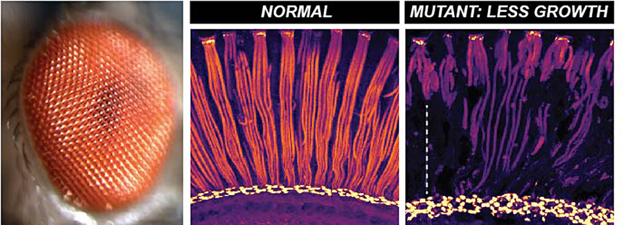
The compound fly eye (left) has multiple, elongated photosensitive compartments (middle, red) that grow during development to span the depth of the retina. Defective growth in mutants (right) results in shorter photosensitive compartments that fail to reach the base of the retina. © Hebbar et al. / MPI-CBG
Restricting an ongoing process is at times crucial – in our daily life as well as in our cells. This is especially important during the development of an organism, when “stop” signals provide an appropriate mechanism to limit growth. A certain shape and size of the developing cell, tissue, organ, or organism emerges due to some kind of restriction of an ongoing growth process.
The research group of Elisabeth Knust at the Max Planck Institute of Molecular Cell Biology and Genetics (MPI-CBG) in Dresden in collaboration with the research group of Andrej Shevchenko, also located at the MPI-CBG, studied the growth of the photosensitive (light-absorbing) compartment in the retina of the fruit fly Drosophila melanogaster. The second-half of eye development is dedicated to its growth. For example, the photosensitive compartment needs to grow more than two-fold in length to span the retinal depth. This compartment in the fly eye is similar in many ways to the photosensitive compartment of our human eyes, and its correct size is crucial for its function, namely vision. What are the signals that eventually shape these compartments during development?
In this study published in the Journal of Cell Biology, a particular class of lipids, called hydroxylated sphingolipids, was identified to be responsible for restricting growth. The researchers used fly mutants exhibiting growth defects in their photosensitive compartments. These studies were coupled with a comprehensive analysis of lipids in the eye. The authors explain: “By integrating these tools with a quantitative approach, we were able to identify increased levels of hydroxylated sphingolipids in mutants with shorter photosensitive compartments.” The researchers also found that hydroxylated sphingolipids modulate growth by restricting the amount of the light-sensing protein (Rhodopsin) that is sent to these compartments.
In future studies, it will be important to investigate questions such as:
1. How do other aspects of cell metabolism impact the growth of these specialized compartments?
2. Do similar “stop” signals restrict growth in other tissues? This is important because hydroxylated lipids are also present in specialized compartments of organs such as the intestine.
Sarita Hebbar, Kai Schuhmann, Andrej Shevchenko, Elisabeth Knust: Hydroxylated sphingolipid biosynthesis regulates photoreceptor apical domain morphogenesis. J Cell Biol (2020), 13. October 2020, doi: 10.1083/jcb.201911100